How oxidative stress and autism are actually connected
Oxidative stress in autism is measurable directly in postmortem brain tissue, and it tracks with speech delay, sensory sensitivity and repetitive behavior alike. The mitochondrial half of that story gets far less attention than the antioxidant half, and it changes what a workable protocol actually looks like.
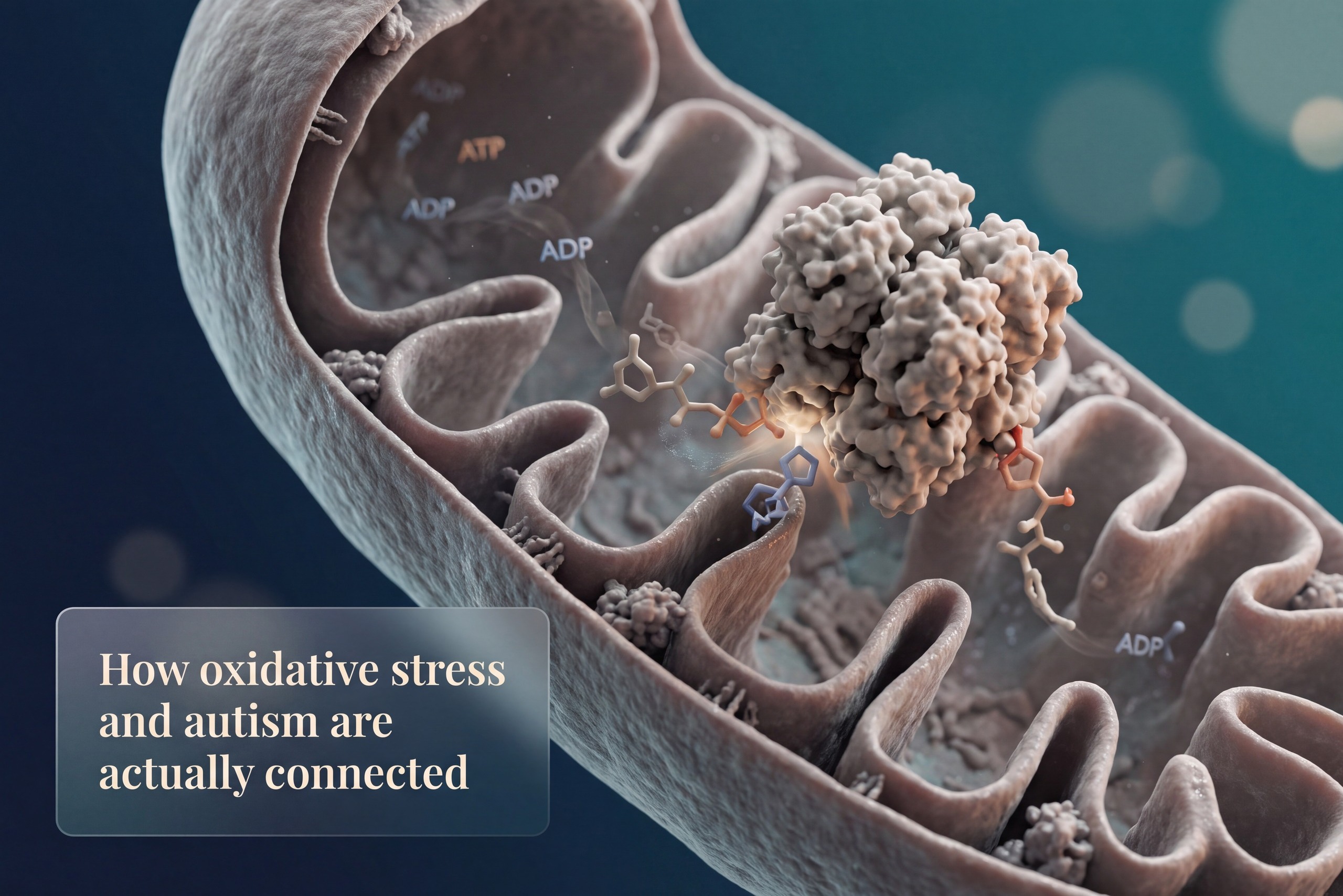
What oxidative stress in autism actually looks like
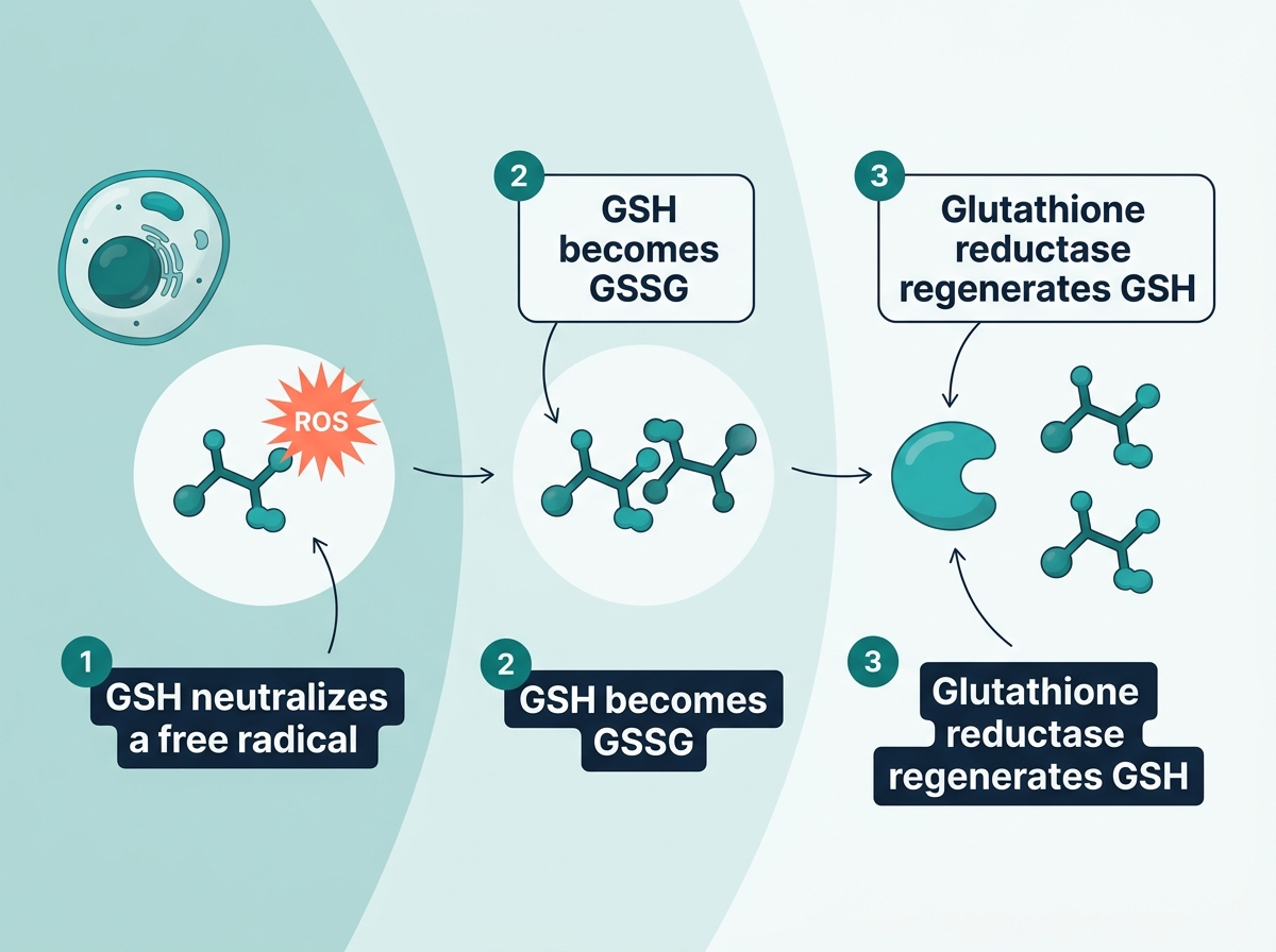
Oxidative stress and autism connect at the level of redox imbalance, the tilt between damaging molecules and the body's cleanup systems. In autism that tilt runs toward damage. Reactive oxygen species, often called free radicals, build up faster than the antioxidant system can clear them. Reduced glutathione, or GSH, is the body's main antioxidant, and its oxidized form, GSSG, is what's left once GSH has already neutralized a threat. The GSH-GSSG ratio is the single number that shows how much active antioxidant capacity remains versus how much has already been spent.
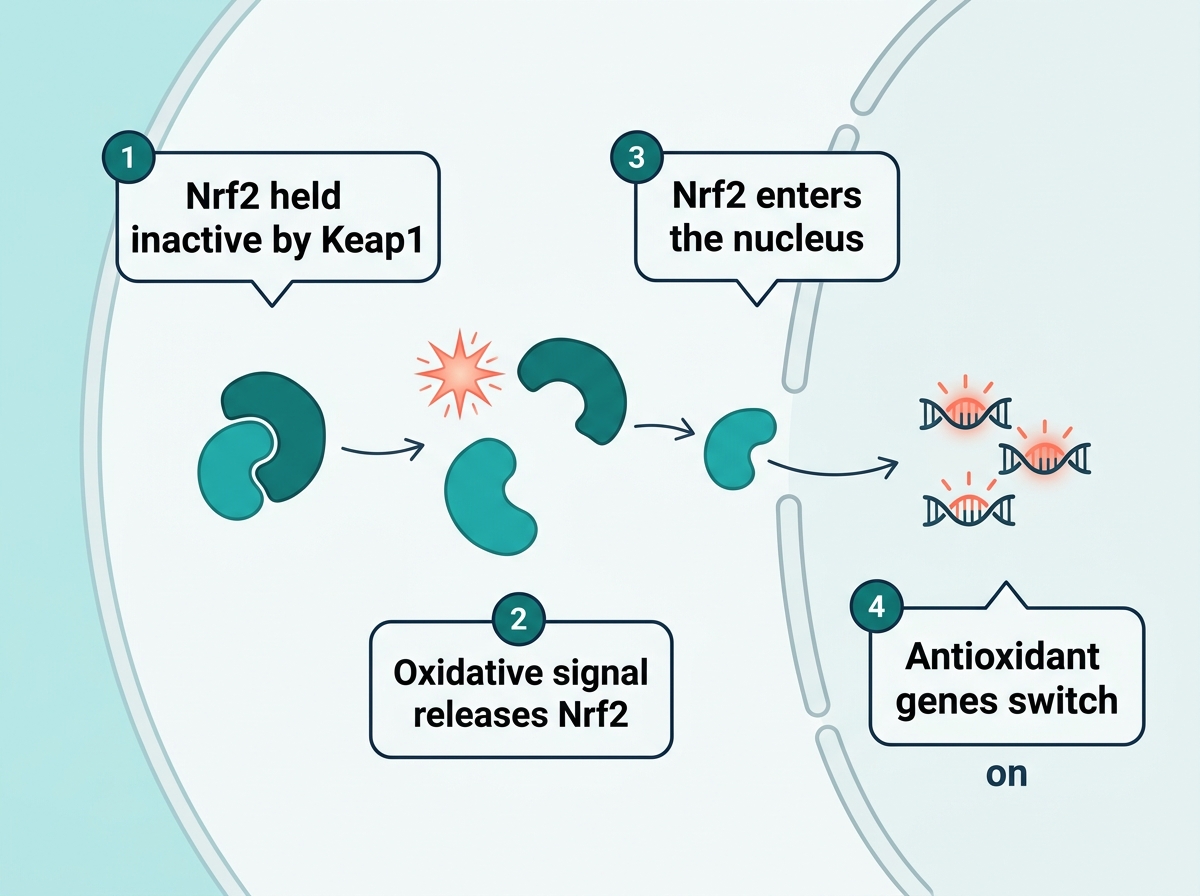
Nrf2 is the molecular switch that turns on antioxidant production when a cell senses trouble, and in autism this switch runs less efficiently.
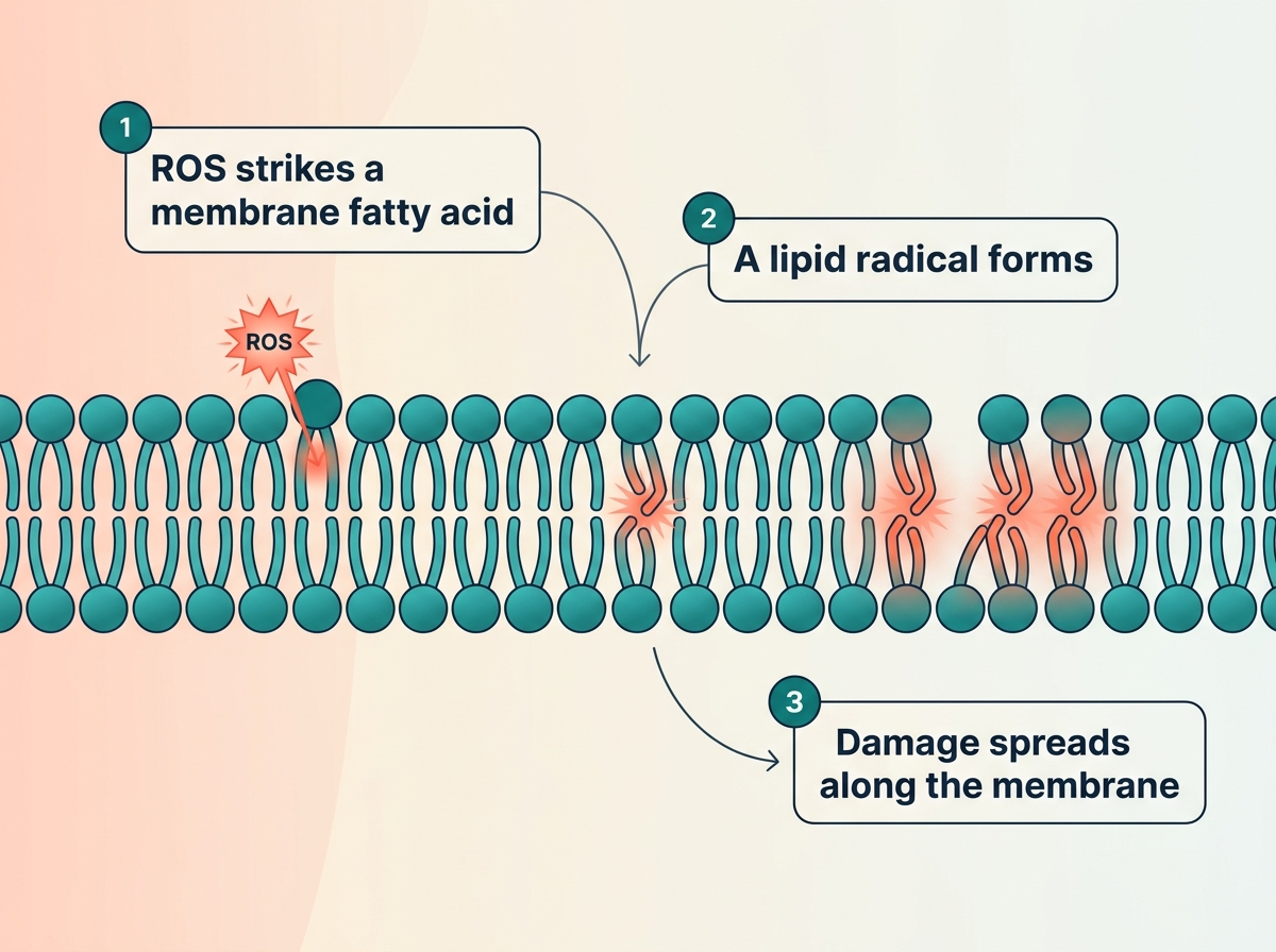
The downstream damage shows up as lipid peroxidation, meaning harm to the fatty membranes surrounding every cell, alongside protein oxidation and DNA damage. Mitochondrial superoxide production adds to the load directly, because mitochondria are the cell's energy batteries, and when they leak, they leak reactive oxygen species straight into the surrounding cytoplasm, which also feeds neuroinflammation over time. A subset of autism cell lines shows measurably greater mitochondrial superoxide output under stress than typically developing cell lines.
Biomarkers of oxidative stress in postmortem brain tissue
Postmortem brain tissue gives the most direct evidence available, since it removes any question about whether a blood marker actually reflects what's happening inside the brain. Researchers measured glutathione directly in autism brain tissue and found GSH reduced by 43% in the cerebellum and 32% in Brodmann area 22, alongside GSSG increased by 18% and 19% in those same regions.
STUDY Postmortem cerebellum and Brodmann area 22 tissue from autistic individuals (n=15) showed GSH reduced 43% and 32%, GSSG increased 18% and 19%, and the GSH-GSSG ratio reduced by 52% and 43% compared with controls (n=12). Rose et al., 2012, Evidence of oxidative damage and inflammation associated with low glutathione redox status in the autism brain, Translational Psychiatry.
Decreased GSH/GSSG redox capacity and increased oxidative stress in the autism brain may have functional consequences in terms of a chronic inflammatory response.
A systematic review and meta-analysis of 87 studies covering more than 9,000 children confirmed this pattern at scale. Oxidative stress markers were consistently abnormal across autism spectrum disorder, a neurodevelopmental disorder where reduced glutathione and a lower GSH-GSSG ratio showed up again and again across dozens of independent research groups.
STUDY A systematic review and meta-analysis of 87 studies covering 9,109 children found consistently abnormal oxidative stress markers in autism spectrum disorder, including reduced GSH, a reduced GSH-GSSG ratio, and elevated GSSG, homocysteine and malondialdehyde. Chen et al., 2021, Oxidative stress marker aberrations in children with autism spectrum disorder, Translational Psychiatry.
Diminished antioxidant capacity shows up in more than a lab report number. The chart below shows how far the GSH-GSSG ratio actually drops in each brain region, and children with less antioxidant reserve tend to score worse on clinical severity ratings, a pattern that keeps recurring across the oxidative stress markers literature in autism spectrum disorder.
Glutathione redox status in postmortem autism brain tissue
Rose et al., 2012, Translational Psychiatry, postmortem brain tissue, autism (n=15) vs control (n=12), shown as % of control mean
Which brain functions and symptoms actually track with oxidative stress
Speech and language impairment tracks measurably with lower antioxidant levels in children with autism spectrum disorder. The prefrontal cortex and temporal lobe, the regions that handle language and social understanding, sit among the tissue most vulnerable to this kind of damage, since both regions carry the high energy demand that makes neurons especially exposed to reactive oxygen species.
The role of oxidative stress in symptom severity
Sensory processing dysfunction has its own specific markers. In a study of 44 autistic children and 40 controls, 8-isoprostane and cysteinyl leukotriene levels predicted sensory sensitivity scores on the Short Sensory Profile scale, with both markers negatively correlated with sensory function.
How strongly oxidative markers track with sensory symptoms
Qasem, Al-Ayadhi and El-Ansary, 2016, Lipids in Health and Disease, 44 autistic children vs 40 controls, correlation with Short Sensory Profile scores shown as absolute strength
Overall autism severity tells a consistent story once four markers are measured together. In an 89-child study built around a diagnosis of autism spectrum disorder under DSM-5 criteria, 8-OHdG, 3-nitrotyrosine and advanced oxidation protein products were all elevated, while the DNA repair enzyme OGG1 was lower, and every one of these four markers correlated with how severe a clinician rated the child's symptoms. Clinicians often reach that rating with tools like the Childhood Autism Rating Scale, and biochemical markers like these now add an objective layer to the screening of autism spectrum disorder alongside them.
STUDY Among 60 children with ASD diagnosed under DSM-5 criteria and 29 controls, 8-OHdG, 3-nitrotyrosine and advanced oxidation protein products were significantly elevated and OGG1 significantly reduced, and all four markers correlated with symptom severity. Ismael and Ismail, 2025, Investigating Oxidative Stress and Impaired DNA Repair Capacity as Diagnostic Biomarkers in Autism Spectrum Disorder, Journal of Molecular Neuroscience.
Repetitive and stereotypic behavior has a newer, more tentative data point behind it. A 2025 mouse study found that plasma glutathione levels predicted the severity of repetitive, stereotypic behavior in younger animals, with fifteen proteins tied to oxidative stress regulation showing the same pattern. This is early-stage mouse research, a signal worth watching well before it becomes a settled finding in children with autism spectrum disorder.
STUDY Plasma glutathione predicted stereotypy severity in younger mice (P=0.0033), with fifteen proteins tied to oxidative stress regulation showing overlapping patterns. Coden et al., 2025, Stereotypy is strongly linked to multiple biomarkers of oxidative stress, PLOS ONE.
The antioxidants usually recommended, and what they cover
N-acetylcysteine, glutathione precursors, CoQ10, vitamin E and polyphenols make up the usual antioxidant toolkit for autism, and each one works the same basic way. They either neutralize reactive oxygen species directly, once damage has already started, or they refill the antioxidant supply the cell has already used up.
A randomized controlled trial gave 46 children a cysteine-rich whey protein, a direct glutathione precursor, over 90 days. Children receiving the active protein showed significant gains on the Vineland Adaptive Behavior Scale composite score, along with improvements across socialization and adaptive behavior domains compared with placebo.
STUDY In a randomized, double-blind trial of 46 children aged 3 to 5 with ASD, cysteine-rich whey protein produced significant gains on the VABS-II composite score (effect size 0.98) plus improvements in adaptive behavior, socialization and internalizing behavior domains. Castejon et al., 2021, Improving Antioxidant Capacity in Children With Autism, Frontiers in Psychiatry.
N-acetylcysteine produced one of the clearer behavioral results in the literature. In a randomized pilot trial, children given NAC saw irritability scores drop from 16.9 at baseline to 7.2 by week 12, compared with a much smaller drop from 14.8 to 13.1 in the placebo group.
Irritability scores before and after N-acetylcysteine
Hardan et al., 2012, Biological Psychiatry, ABC irritability subscale, randomized controlled pilot trial (n=33 randomized)
My son is 6 and was just diagnosed with autism. His pediatrician mentioned his glutathione levels might be low after some bloodwork, but glossed over what that means or what we should do about it. Should I be worried, and is this something a supplement can fix?
Low glutathione on a lab report usually reflects exactly what we've covered here, an antioxidant system running behind on cleanup. It's common in children with autism spectrum disorder, and it's a manageable finding with a clear next step. Precursors like NAC or cysteine-rich whey protein have real trial data behind them, and dosing and timing matter for getting a real result. Glutathione is only half the picture too, the mitochondrial side further down fills in the rest. It deserves a proper, testing-based look.
| Cleans up damage after it happens | Prevents damage before it starts |
|---|---|
| NAC, glutathione precursors, CoQ10, vitamin E, polyphenols | Mitochondrial creatine kinase, recycling ADP at the inner membrane |
| Neutralizes reactive oxygen species once they've already formed | Stops reactive oxygen species from forming in the first place |
The mitochondrial route, how creatine kinase prevents ROS at the source
Mitochondrial creatine kinase is an enzyme that sits at the inner mitochondrial membrane and handles ADP recycling, meaning it helps regenerate the cell's energy currency as fast as the cell burns through it. That recycling step goes a full level deeper than the antioxidants in the previous section. It prevents reactive oxygen species from forming in the first place, at the earliest possible point in the chain.
The phosphocreatine-to-creatine ratio determines how strong this effect actually is. Researchers testing the mechanism directly on brain tissue, including synaptosomes, isolated mitochondria and microsomes, found the antioxidant effect held up consistently across multiple different damage models.
STUDY Mitochondrial creatine kinase activity, through ADP recycling, prevented reactive oxygen species generation across synaptosomes, isolated mitochondria and microsomes under multiple oxidative damage models. Meyer et al., 2006, Mitochondrial Creatine Kinase Activity Prevents Reactive Oxygen Species Generation, Journal of Biological Chemistry.
What I see in practice
In my own clinical work, children who start creatine under the specific protocol I lay out in the book, alongside proper testing, tend to show changes that fit this mechanism closely. Parents report better muscle tone within weeks, more sustained eye contact, and noticeably faster response to simple commands. I've seen this pattern repeat often enough across cases that it stopped looking like coincidence to me a long time ago, even though it still needs the kind of controlled trials that autism research hasn't run yet.
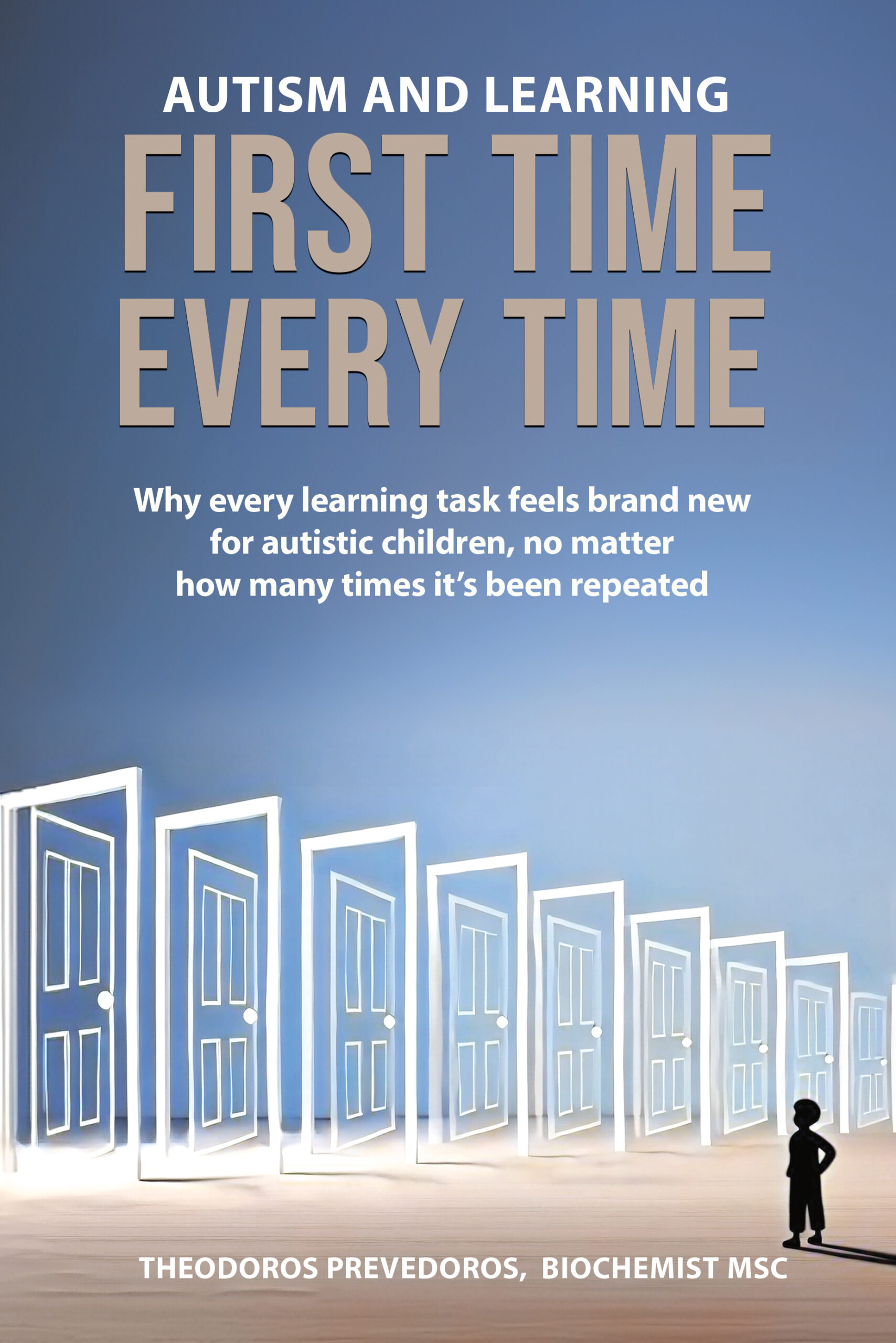
See the full protocol
This is the exact mechanism the creatine protocol in Autism and Learning is built around. If muscle tone, eye contact or responsiveness are part of what you're watching for, the book walks through how to structure it safely. Buy the bookWhy this connects back to the mitochondrial dysfunction already documented in autism
31P-MRS is a type of brain scan that measures energy molecules directly, without a biopsy, by reading the signal from phosphorus atoms inside living tissue. The first study to apply it in autism found evidence of undersynthesis and increased breakdown of brain membrane phospholipids, a signature of disrupted energy and membrane metabolism spread across several markers at once.
STUDY The earliest phosphorus-MRS study in autism found evidence for undersynthesis and increased degradation of brain membrane phospholipids. Minshew et al., 1993, A preliminary 31P MRS study of autism, Biological Psychiatry.
A later, smaller follow-up extended the same scanning approach from muscle into brain tissue in children with autism spectrum disorder and found the same directional pattern, phosphocreatine trends consistent with lower available energy reserves. The antioxidant story from the earlier sections and this energy story are the same problem, viewed from two different angles, and creatine sits exactly at the intersection, one mechanism touching both at once.
What this means in practice
The creatine kinase mechanism is decades-established science, already studied extensively in neuroscience and mitochondrial dysfunction research. Parkinson's disease and other neurodegenerative conditions have used it for years, where creatine and phosphocreatine measurably reduce oxidative stress and protect neurons in laboratory models.
STUDY Both creatine and phosphocreatine reduced oxidative stress and provided measurable neuroprotection in an in vitro Parkinson's disease model. Cunha et al., 2014, Both Creatine and Its Product Phosphocreatine Reduce Oxidative Stress and Afford Neuroprotection, ASN Neuro.
Autism is the frontier where researchers are only beginning to apply this established mechanism at scale, even though the biochemistry connecting oxidative stress and autism, mitochondrial energy production above all, is the same biochemistry described throughout this article. That gap is exactly the opportunity.
I've read that creatine helps with muscles in athletes, but I saw somewhere online that it might help with brain fog or low muscle tone in autistic kids too. Is there any real science behind that or is it just another supplement fad parents are trying out of desperation?
It's a fair question, because most of what circulates online about creatine has nothing to back it up. This case is different. Creatine kinase's role in preventing oxidative damage is well-established neuroscience, tested directly on brain tissue and already used in Parkinson's research for years. That mechanism is solid. Autism research still needs trials at that same scale. Clinically, a structured protocol closes part of that gap already, built on proper testing and individualized dosing.
A related piece on this site walks through exactly what a starved mitochondrial energy supply looks like in an autistic brain, and it converges on the same biochemistry from a different angle. Reading the mechanism here alongside the case-level detail there tends to make the whole picture click into place, which is exactly the walkthrough the book builds on chapter by chapter.
Ready to see the full mitochondrial protocol?
Autism and Learning: First Time Every Time walks through the creatine, antioxidant and testing protocol behind everything in this article, in the order I actually use it with families.
Frequently asked questions
The classic panel includes lower GSH, higher GSSG, a reduced GSH-GSSG ratio, elevated 8-OHdG, 3-nitrotyrosine and advanced oxidation protein products, and reduced OGG1. One creatine-specific marker belongs on this list too, guanidinoacetate. It builds up when creatine synthesis is inefficient, and it directly increases free radical formation while lowering brain antioxidant defenses, which is part of why creatine status and oxidative stress are linked biochemically.
The prefrontal cortex and temporal lobe carry the heaviest energy demand of any language-related tissue, which makes their neurons unusually exposed to reactive oxygen species. One specific pathway runs through lipid peroxidation of the fatty membranes that insulate nerve fibers, including the white-matter tracts that connect language regions to each other. Damage there slows the exact signal transmission speech processing depends on, which is a plausible mechanistic reason lower antioxidant status tracks with more communication difficulty in the research.
Mitochondria generate the energy every neuron runs on, and multiple lines of evidence, from brain scans to cell studies, point to that energy supply running short in a meaningful subset of autistic children. The gut microbiome adds another route into the same problem. Propionic acid, a short-chain fatty acid produced by gut bacteria, directly alters mitochondrial energy output in autism-derived cells, and its effects turn harmful specifically once reactive oxygen species are already elevated, tying the microbiome, mitochondria and oxidative stress into one connected system.