Glutathione deficiency and autism, and how it is tested
Glutathione hands an electron to oxidants and gets rebuilt for the next round. That makes it the body's main cleanup and recycling system. Glutathione deficiency and autism connect through blood and brain studies. Those studies also find a lower ratio of working to used glutathione in children with autism. The amino acid glycine, shared with creatine, sets how fast the cycle restarts. Together, they form one supply chain for cell cleanup and cell energy.
What does glutathione actually do in the body?
Glutathione hands an electron to a free radical or another reactive oxygen species and disarms it. The cell then rebuilds the used molecule for the next round. It works like a reusable sponge, so the same unit gets wrung out and used again. Cells build it from three amino acids, glutamate, cysteine and glycine, joined in one short chain. The middle unit carries a free sulfur group (a thiol) that does the electron handoff.
Each handoff turns reduced glutathione (GSH) into oxidized glutathione (GSSG), two units bridged by a sulfur-sulfur bond. Then the enzyme glutathione reductase, powered by NADPH, snaps that bridge and returns two working units. Under normal conditions, more than 90% of the cellular pool stays in the reduced form.
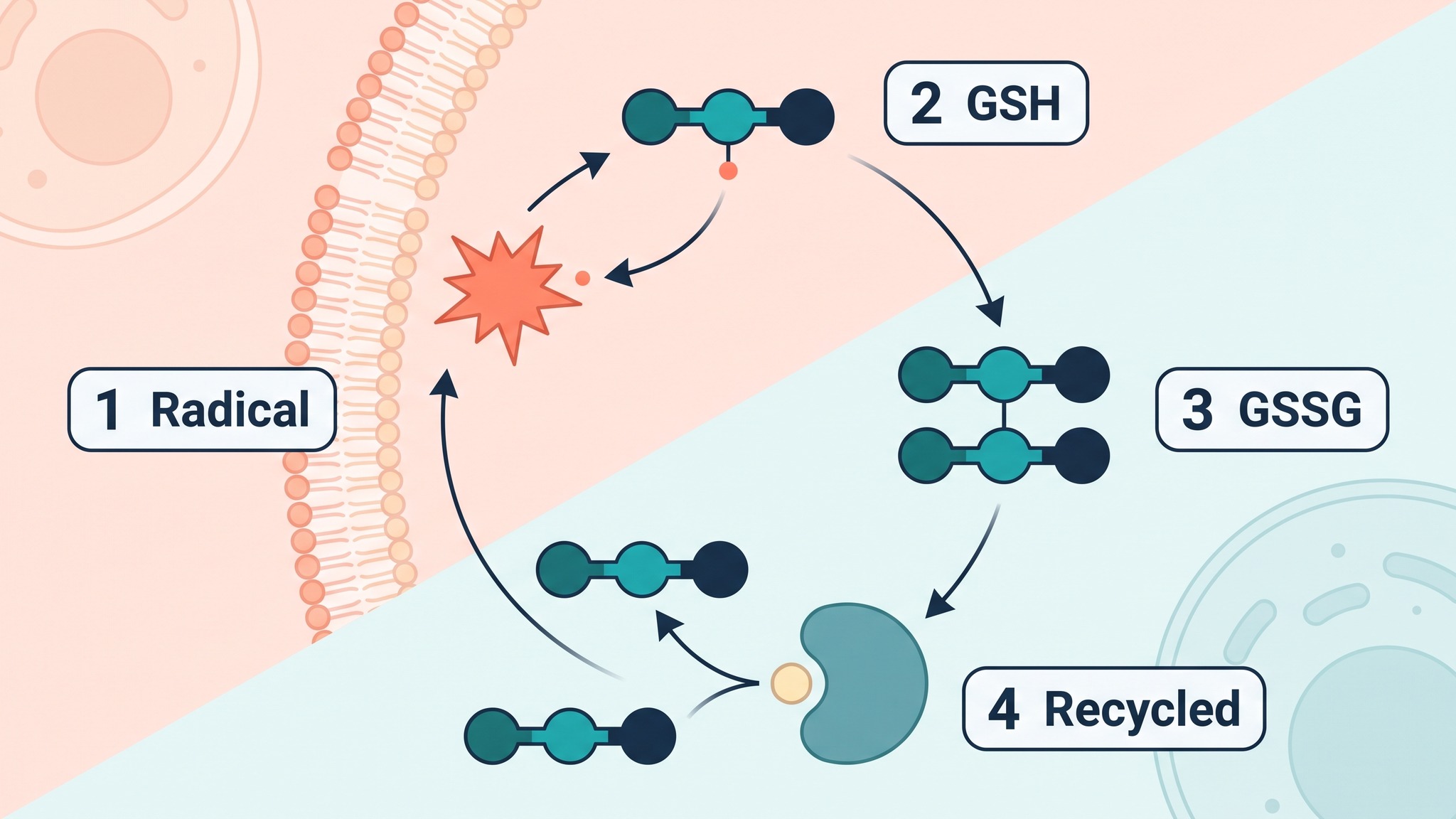
Glutathione is an antioxidant with a built-in recycling route, and two enzyme families put it to work. Glutathione peroxidases, for instance, use it to turn hydrogen peroxide into water. Each glutathione S-transferase enzyme attaches it to a foreign chemical (a xenobiotic) so the body can excrete that chemical.
Its intracellular concentration runs from 0.5 to 10 millimolar (mM), so cells keep a large working pool. Oxidative stress is the state in which oxidants arrive faster than this cycle can clear them. The balance between the two forms is the cell's redox balance, and most laboratory tests try to capture it.
How is glutathione deficiency diagnosed?
Glutathione deficiency is diagnosed from whole blood, where a lab measures GSH. Some panels add its ratio to the oxidized form. Red blood cells hold nearly all of the blood's glutathione, so a whole-blood tube reflects the red-cell pool. Red-cell glutathione is the biomarker most labs report. Each lab publishes its own reference range. So a result is read against the range from the same method.
Where in the blood does glutathione sit?
Red blood cells carry glutathione at millimolar concentrations, while plasma holds only 0.1 to 20 micromolar (µM). Plasma glutathione also has a half-life of 1.6 minutes, so a plasma reading captures one moment. Sample handling then decides the result. Air exposure after the draw oxidizes glutathione in the tube. That creates an abnormality the body never had.
STUDY Acid deproteinization produced 30 to 150 µmol/L of the oxidized form, against a true 2 to 6 µmol/L. Published blood values span 1 to more than 500 µmol/L. Rossi et al., 2002, Blood Glutathione Disulfide: In Vivo Factor or in Vitro Artifact?, Clinical Chemistry.
Adding N-ethylmaleimide (NEM) at the moment of collection locks glutathione in place and prevents that artifact.
What do the most common tests offer and where do they fall short?
Eight tests dominate the market, and each measures a different piece of the glutathione picture.
| Test | What it offers and where it falls short |
|---|---|
| Labcorp 007700, total glutathione (whole blood, LC-MS/MS) | Orderable in the US, stable for 14 days at room temperature. Total glutathione only, with an investigational label and no reference range. |
| Mayo glutathione, blood (DTNB spectrophotometry) | Reference range from 12 months of age, 46.9 to 90.1 mg/dL of red cells. A white cell count above 20 billion per liter raises the result by up to 25%. A recent transfusion also makes it unreliable. |
| Consumer dried blood spot (LC-MS/MS) | Finger-prick at home, reporting both forms and the ratio in 3 to 5 business days. The vendor's page says global clinical cutoffs are still undefined. |
| Research finger-stick assay | Uses 10 to 20 µL of blood with NEM added at once. It matches HPLC at r = 0.9746 for the reduced form and 0.9693 for the oxidized form. |
| Plasma glutathione and ratio by HPLC | Used in the blood studies below. Plasma sits between 0.1 and 20 µM, and acid handling inflates the reading. |
| Urine pyroglutamic acid | Flags strain on the glutathione cycle after acetaminophen, alcohol, high-fructose diets or fasting beyond 24 hours. It reads the cycle indirectly, so the cause stays hard to assign. |
| 8-OHdG in urine or serum | Tracks oxidative DNA damage. ELISA kits read 41.6 pmol/mL higher on average than LC-MS/MS, and urea cross-reacts with the antibody. |
| GST genotyping (GSTM1, GSTT1, GSTP1, GSTA1) | Inherited traits that diet and supplements leave unchanged. In 113 affected children and 114 controls, GSTP1 IleIle with active GSTM1 gave an odds ratio of 11.088 (95% CI 1.745 to 70.456). |
Which questions should you ask the lab?
Five questions decide whether a glutathione result can be trusted. The lab can answer each one before the blood draw.
- Is the sample whole blood or plasma, and which one does the reference range describe?
- Was NEM or another stabilizer added at collection?
- Which method produced the number, and does it come with a pediatric reference range?
- How long did the sample travel, and at what temperature?
- Has the child had a recent transfusion or a high white cell count?
What links glutathione deficiency and autism in blood and brain research?
Glutathione in autism spectrum disorder (ASD) has been studied in blood, brain tissue and genes. Overall, the results converge on a lower ratio of working to used glutathione. A meta-analysis of 87 studies covered 4,928 cases and 4,181 controls. It found lower GSH, lower total glutathione and a lower ratio, plus a higher level of the oxidized form. So this neurodevelopmental condition shows a measurable shift in glutathione status.
- GSH:GSSG ratio
- The amount of glutathione ready to work (the reduced form) divided by the amount already used (the oxidized form). A high ratio means the cleanup keeps pace with demand.
What do blood studies of children with autism show?
In blood, these children carry less glutathione and a smaller share of it in working form.
STUDY The total-to-oxidized ratio was 8.6 in 20 autistic children against 25.5 in 33 controls. Total glutathione was 4.1 against 7.6 µmol/L. James et al., 2004, Metabolic biomarkers of increased oxidative stress and impaired methylation capacity in children with autism, American Journal of Clinical Nutrition.
Blood markers in affected children as a percent of controls
James et al., 2004. Control group mean = 100%. Percentages computed from the reported group means (20 children, 33 controls).
Alongside glutathione, the methyl donor SAM stood at 78% of the control value. The SAM to SAH ratio, a marker of methylation, stood at 56%. Pooled data from 29 articles show the same direction. There, the working form sits 27% lower and the oxidized form 45% higher.
Pooled differences in blood markers, cases versus controls
Frustaci et al., 2012, 29 articles pooled. Bar length is proportional to the size of the difference. The sign is in the label.
Young children show the pattern in plasma too, alongside lower Nrf2. Nrf2 is the transcription factor that switches on antioxidant genes. In 23 children aged 1.6 to 4.0 and 21 controls, Nrf2 tracked with glutathione and the ratio. However, another study of 49 children and 31 controls found equal glutathione in both groups (p = 0.30).
What is the role of glutathione in brain function?
Brain cells hold 1 to 2 mM of glutathione. Astrocytes (star-shaped support cells) store about 8 mM and then export roughly 10% of it per hour. The enzyme γ-glutamyl transferase on neighboring cells splits the exported glutathione into building blocks. Then neurons take up those fragments to build their own.
STUDY Postmortem tissue from 15 people with autism held 43% less GSH in the cerebellum than tissue from 12 controls. Brodmann area 22 (BA22), a region for hearing and language, held 32% less. Rose et al., 2012, Evidence of oxidative damage and inflammation associated with low glutathione redox status in the autism brain, Translational Psychiatry.
Glutathione markers in postmortem brain tissue, cases versus controls
Rose et al., 2012, 15 cases and 12 controls. Bar height is proportional to the size of the change. The sign is in the label.
Also, in frontal cortex, the gene for Nrf2 (NFE2L2) sat 30.5% lower in 7 cases than in 8 controls.
Which symptoms and functions track with glutathione status?
In autism spectrum disorder, markers of glutathione status track with severity, sensory processing and adaptive function across several studies.
In 55 children with ASD and 44 controls, plasma glutathione ran lower (p below 0.0001). The oxidized form ran higher, and it was a significant variable in all three severity-scale models (adjusted R² 0.15 to 0.24). Similarly, in 89 children, oxidative damage markers tracked severity. These included 8-OHdG (p = 0.029), 3-nitrotyrosine (p = 0.0043) and AOPP, a protein damage marker (p = 0.046). Lower OGG1, a DNA repair enzyme, also linked to severe symptoms (p = 0.0001).
Next, sensory processing follows the same direction. Among 44 children with ASD and 40 controls, higher 8-isoprostane went with lower Short Sensory Profile scores (r = −0.517). This marker reflects lipid oxidation, and lower scores mean more sensory difficulty. Also, cysteinyl leukotrienes, which are inflammatory lipids, reached r = −0.615.
For speech and language, the nearest data come from the temporal lobe and from genes. BA22, a hearing and language region, held 32% less glutathione in that postmortem study. In 113 children with ASD, the GSTA1 CC genotype went with less impairment in non-verbal communication (β = −0.232). Active GSTM1 went with higher adaptive scores on the Vineland scale (β = 0.160).
Finally, repetitive behavior enters the picture in mice. Depleting glutathione with buthionine sulfoximine exaggerated repetitive behavior in BTBR mice, an inbred strain that models autism-like behavior.
Does swallowing glutathione raise blood levels, and which routes work best?
Swallowing glutathione raises blood levels when the dose is taken daily for weeks. A single large dose, however, leaves plasma unchanged. Route, dose and duration decide the result. The ways to increase glutathione fall into three groups. These are the finished compound, its raw materials and the switch that turns on its production.
What happens to oral glutathione in the gut?
Enzymes in the gut wall and liver cut oral glutathione into its building blocks before it reaches the blood.
STUDY Seven healthy volunteers took one 3 g oral dose. Plasma glutathione and its building blocks stayed unchanged for 270 minutes. The dose equaled 0.15 mmol/kg, and baseline glutathione was 6.2 µmol/L. Witschi et al., 1992, The systemic availability of oral glutathione, European Journal of Clinical Pharmacology.
Cells import the fragments and rebuild glutathione inside, which is why daily dosing raises levels over weeks.
Which delivery forms raise levels best?
Daily dosing and newer formulations raise blood glutathione, and the size of the rise depends on the form. In a 6-month trial of 54 non-smoking adults, 1,000 mg a day raised glutathione by 30% to 35%. The rise also appeared in red cells, plasma and lymphocytes. Liposomal glutathione raised whole-blood glutathione by 40% in 12 adults after 4 weeks. In 14 adults, one micellar dose gave 2.49 times the exposure of standard 500 mg glutathione (p = 0.0064).
Rise in blood glutathione by delivery route
Single 3 g dose in plasma (Witschi 1992). Oral 1,000 mg a day in red cells, plasma and lymphocytes (Richie 2015). Liposomal 500 to 1,000 mg a day in whole blood (Sinha 2018). Oral or transdermal in whole blood, a nonsignificant rise (Kern 2011). Bar length is proportional to the upper value out of 40%.
Which forms have been tested in children?
Three small studies have tested oral, transdermal and sublingual glutathione in children with ASD.
STUDY Two groups of 13 children with ASD were treated for 8 weeks. An oral form raised plasma glutathione significantly, while a transdermal form raised it 11% (nonsignificant). Whole-blood glutathione also rose 9% to 12% in both. Kern et al., 2011, A clinical trial of glutathione supplementation in autism spectrum disorders, Medical Science Monitor.
An open-label study of 25 participants aged 7 to 18 with ASD tested sublingual glutathione for 6 weeks. The reduced form, the ratio and total antioxidant capacity improved, and the authors reported behavioral gains. A 12-week trial gave 6 children (mean age 14.7) oral glutathione at 500 to 1,000 mg twice daily. The level normalized in the 3 children who started low, and 4 of the 6 had an upset stomach. Each form rests on its own small study, so a direct comparison in children with ASD remains open.
Do NAC and other precursors work better?
N-acetylcysteine (NAC) is a precursor, a raw material the cell uses to build its own glutathione. Oral NAC reaches the blood at 4.0% in its reduced form and 9.1% in total. A meta-analysis of 5 randomized trials tested NAC on the Aberrant Behavior Checklist. It improved the irritability subscale by 4.07 points over placebo (95% CI 1.13 to 7.04). Sulforaphane, a third route, works through Nrf2 modulation. One trial of 29 males aged 13 to 27 tested it for 18 weeks. Checklist scores fell 34% (p below 0.001), against under 3.3% on placebo.
My son is 7, has autism and eats almost no meat or eggs. I bought liposomal glutathione powder after a parent group recommended it. Does it actually reach his blood, or should I test him first?
Liposomal glutathione raised whole-blood levels by 40% in adults after four weeks, so the form is worth a look. With a low-meat diet, though, your son's protein intake sets how fast his red cells rebuild glutathione. Glycine is the first amino acid to run short when protein drops. A baseline whole-blood test with proper handling gives you a number to compare against. Then start one change with his clinician and retest under the same conditions.

Help your child learn each skill first time, every time
Once the biology is on the table, the next question is daily life. Autism and Learning: First time Every time shows parents and educators how every child can learn. Buy the bookHow does glycine connect glutathione and creatine?
Glycine is a raw material for both glutathione and creatine. So the two pathways draw on one amino acid pool. Glutathione synthase adds it in the final step of glutathione synthesis. The enzyme AGAT (arginine:glycine amidinotransferase) uses it in the first step of creatine synthesis. Glutathione metabolism therefore shares this amino acid with the pathway that builds creatine. Our article on creatine and brain energy covers the supply chain from AGAT and GAMT to the SLC6A8 transporter.
Glycine supply sets the pace of glutathione synthesis in red cells. In 8 older adults, red-cell glycine sat at 218.0 µmol/L against 486.7 in 8 young adults. Red-cell glutathione was 1.12 against 2.08 mmol/L, and synthesis ran at 45.80% against 83.14% per day. After two weeks of glycine supplementation together with a sulfur-containing amino acid, glutathione rose by 94.6%.
STUDY Cutting protein from 1.13 to 0.75 g/kg a day slowed red-cell glutathione synthesis in 12 healthy adults. The effect appeared within 3 days, and glycine was the limiting amino acid. Jackson et al., 2004, Synthesis of erythrocyte glutathione in healthy adults consuming the safe amount of dietary protein, American Journal of Clinical Nutrition.
For example, a 70 kg adult drops from 79 g to 52.5 g of protein a day.
How much glycine does creatine synthesis use?
Each creatine unit built in the body consumes a whole glycine. Dietary intake data put glycine at 59.6 mg/kg per day. Creatine made from precursors reaches 41.9 mg/kg per day. Converted to its glycine equivalent, that is about 24 mg/kg per day, roughly 40% of the dietary intake.
However, since dietary creatine is only provided in animal products, principally in meat and fish, virtually all of the creatine loss in vegetarians must be replaced via endogenous synthesis.
So the shared pool matters most where supply falls short, as in low-protein diets, plant-based diets and older age.
What causes low glutathione levels?
Glutathione runs low when demand outpaces the supply of raw materials or when recycling slows. Supply comes first, since dietary protein sets the raw materials available. Demand comes next, because a heavy oxidant load uses glutathione faster. Recycling depends on glutathione reductase and NADPH. Genes add a fixed layer. In 113 affected children and 114 controls, active GSTM1 carried an odds ratio of 0.554 for ASD. The 95% confidence interval ran from 0.313 to 0.983.
How can creatine supplementation ease the load on this shared glycine?
Creatine feeds back on AGAT and slows the enzyme's own output, so less glycine flows into the creatine pathway. In human HAP1 cells, creatine suppressed AGAT expression in a dose-dependent way. Half-maximal suppression came at 1 to 2 mM of intracellular creatine.
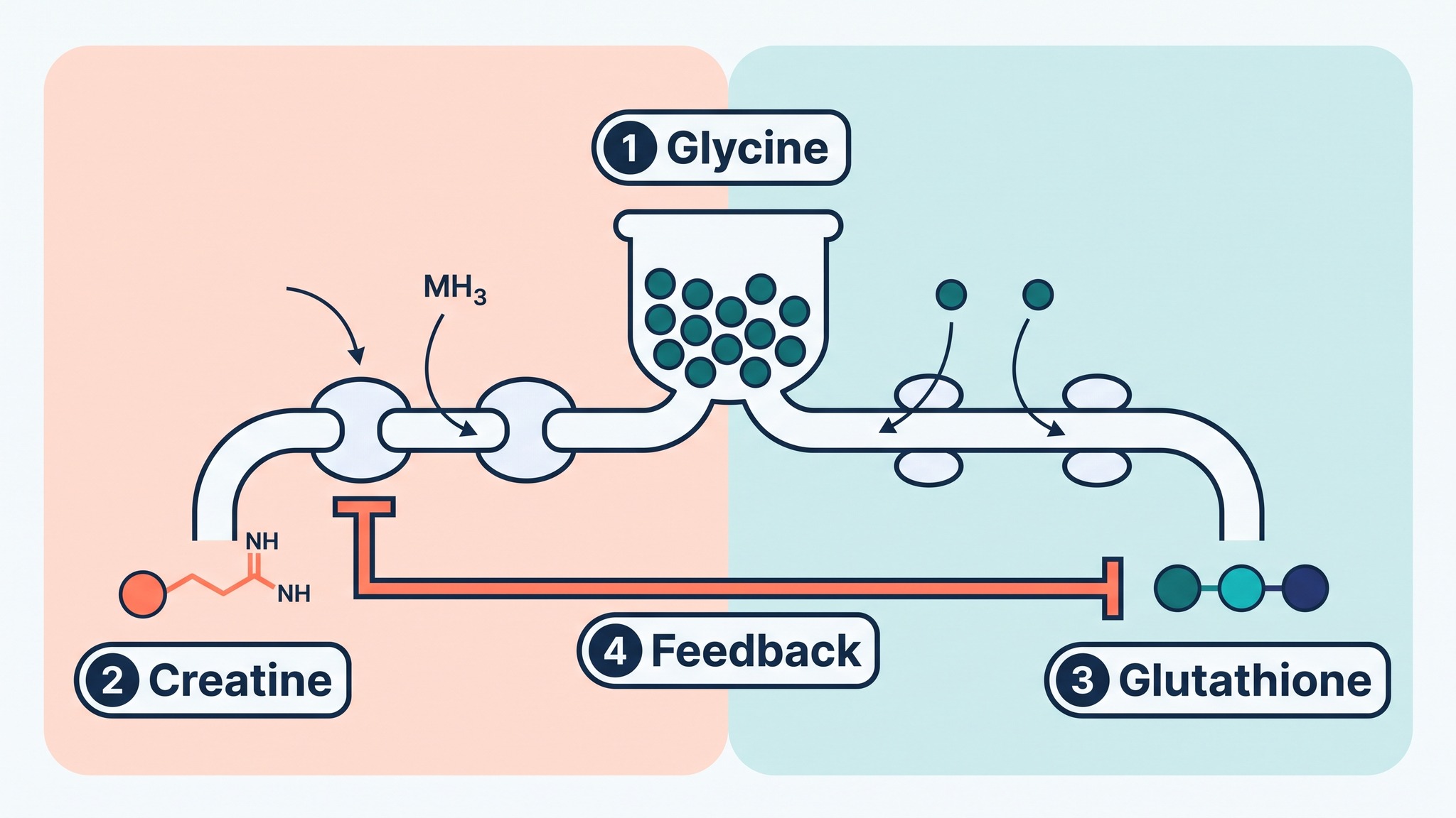
Mitochondrial creatine kinase recycles ADP. In isolated rat brain and liver mitochondria, this recycling lowered reactive oxygen species output. So fewer reactive oxygen species leave glutathione fewer oxidants to clear.
Hybrid striped bass on a glycine-poor diet received 1% and 2% of the amino acid. Creatine and glutathione rose together in the Phase-I liver, with glutathione up 8% and 6%. Muscle and kidney showed no glutathione change, so the link is tissue-specific.
Our article on creatine for autistic people covers genetic deficiencies in AGAT, GAMT and the creatine transporter. The combined question, whether creatine supplementation frees glycine for glutathione in children with ASD, awaits its first human trial. Each half of the mechanism is documented, which makes the question testable.
Why is it so hard to prove that raising glutathione works?
Glutathione turns over so fast that a single blood draw catches one moment of a moving system. Plasma glutathione has a half-life of 1.6 minutes. Red cells rebuild their glutathione at 83.14% per day in young adults. Also, protein intake shifts that rebuilding within 3 days.
Does blood mirror the brain?
Blood serves as a stand-in for the brain, and the two disagree. In 31 adults with autism and 40 controls, magnetic resonance spectroscopy found equal glutathione in two brain regions. Postmortem tissue, by contrast, showed 43% and 32% less. A study of 24 adults with ASD and 18 controls measured 9.6% less glutathione in the anterior cingulate cortex. The values were 1.79 against 1.98 mM (p = 0.076).
Gastrointestinal symptoms add variation. They are 4.42 times as likely in children with ASD. The odds are 3.63 for diarrhea, 3.86 for constipation and 2.45 for abdominal pain.
What do the trials measure so far?
Most clinical trials so far measure blood chemistry. The groups are small. One case series enrolled 6 children, another 13 per group, and an open-label study 25.
My daughter's whole-blood glutathione came back low, so we started NAC and glycine. Eight weeks later the retest is almost the same, but her teachers say she is less irritable. Is the retest wrong, or is the supplement not working?
Both readings can be right, because they measure different things. A blood retest reflects recent glutathione supply. A sample without an immediate stabilizer can also drift on its own. Her teachers' report is a behavior outcome, the same kind of outcome the trials use. For the next draw, keep the same lab, tube and time of day. Then write down what her teachers see in the classroom.
What does this mean in practice?
For families weighing glutathione deficiency and autism, the sequence is simple. Measure once with proper handling, change one thing, then retest under identical conditions.
A whole-blood test with immediate stabilization gives the cleanest baseline. The same lab, tube and time of day then keep the retest comparable. Protein intake shifts red-cell glutathione synthesis within days. So the diet on the day of the draw belongs in the notes. Track function too, meaning sleep, mood, sensory reactions and daily skills, because those are the outcomes the trials use.
At medcasestudies.com, we read a glutathione result next to diet, gut symptoms and developmental history.
The skills regression article on this site follows the same axis. It pairs altered glutathione redox markers with creatine and mitochondrial reserve. Read together, the two pieces connect cell energy with cell cleanup.
The practical goal is a child who uses each learned skill reliably, day after day.
Help your child learn in their own way
Autism and Learning: First time Every time is a practical guide for parents and educators on how every child can learn in their own way.
Frequently asked questions
Oxidative stress is a state in which oxidants are produced faster than the body's antioxidant systems can clear them. Meta-analyses report lower glutathione and a lower ratio of working to used glutathione. Oxidative damage markers such as 8-OHdG and 3-nitrotyrosine also track with severity in 89 children.
A research method can. It uses 10 to 20 µL of blood. The protocol adds N-ethylmaleimide (NEM) immediately and holds the sample for 2 minutes. The result matches HPLC at r = 0.9746 for the reduced form and 0.9693 for the oxidized form. Consumer dried blood spot kits also use finger-prick sampling. Their own pages state that global clinical cutoffs are still undefined.
Daily oral dosing raised levels in healthy adults. Glutathione ran 30% to 35% higher after 6 months of 1,000 mg a day. A single 3 g dose, by contrast, left plasma glutathione unchanged over 270 minutes. In children with ASD, an 8-week oral form raised plasma glutathione significantly in 13 children.
It is a precursor, N-acetylcysteine, that supplies the raw material for the cell's own glutathione. In 6 volunteers, a 400 mg oral dose reached the blood at 4.0% in its reduced form. Total absorption came to 9.1%.