Oxidative stress in autism spectrum and brain energy
Oxidative stress can affect many parts of the autistic brain. It can add pressure to sensory overload, uneven speech, and fatigue. Low glutathione, weak mitochondrial energy, and altered creatine handling give clinicians practical ways to investigate its role in an individual autistic child.
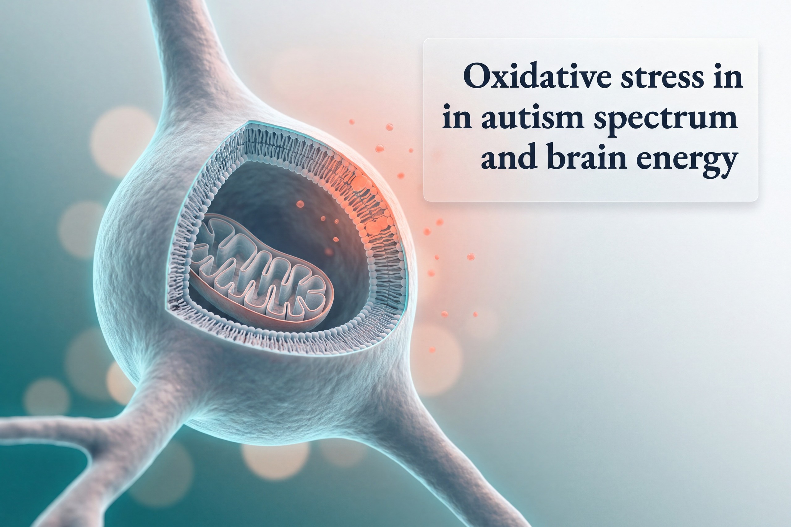
What oxidative stress in autism spectrum looks like in the lab
This laboratory pattern means that reactive oxygen species, often called ROS, are using up antioxidant reserves faster than cells can rebuild them. For a parent, that can make a laboratory report less mysterious. Low reduced glutathione, or GSH, means less of the cell's main antioxidant is available. Higher oxidized glutathione, or GSSG, means more of that reserve has already been used. The GSH/GSSG ratio shows the balance between active protection and spent antioxidant capacity.
The strongest direct brain evidence comes from frozen postmortem tissue and complements blood findings. In cerebellum and Brodmann area 22, a temporal-cortex region involved in speech processing, researchers measured lower GSH and lower GSH/GSSG alongside higher 3-nitrotyrosine and 8-oxo-deoxyguanosine. Those markers reflect protein oxidation and oxidative DNA damage. Lower aconitase activity also pointed to greater mitochondrial superoxide exposure. The same samples linked redox capacity with oxidative injury.
From redox imbalance to cellular damage
Postmortem brain redox differences from controls
Absolute percentage differences versus controls in postmortem autism cerebellum and temporal cortex. Chauhan et al., 2012, DOI 10.1007/s11064-012-0775-4.
How glutathione keeps a neuron’s membrane usable
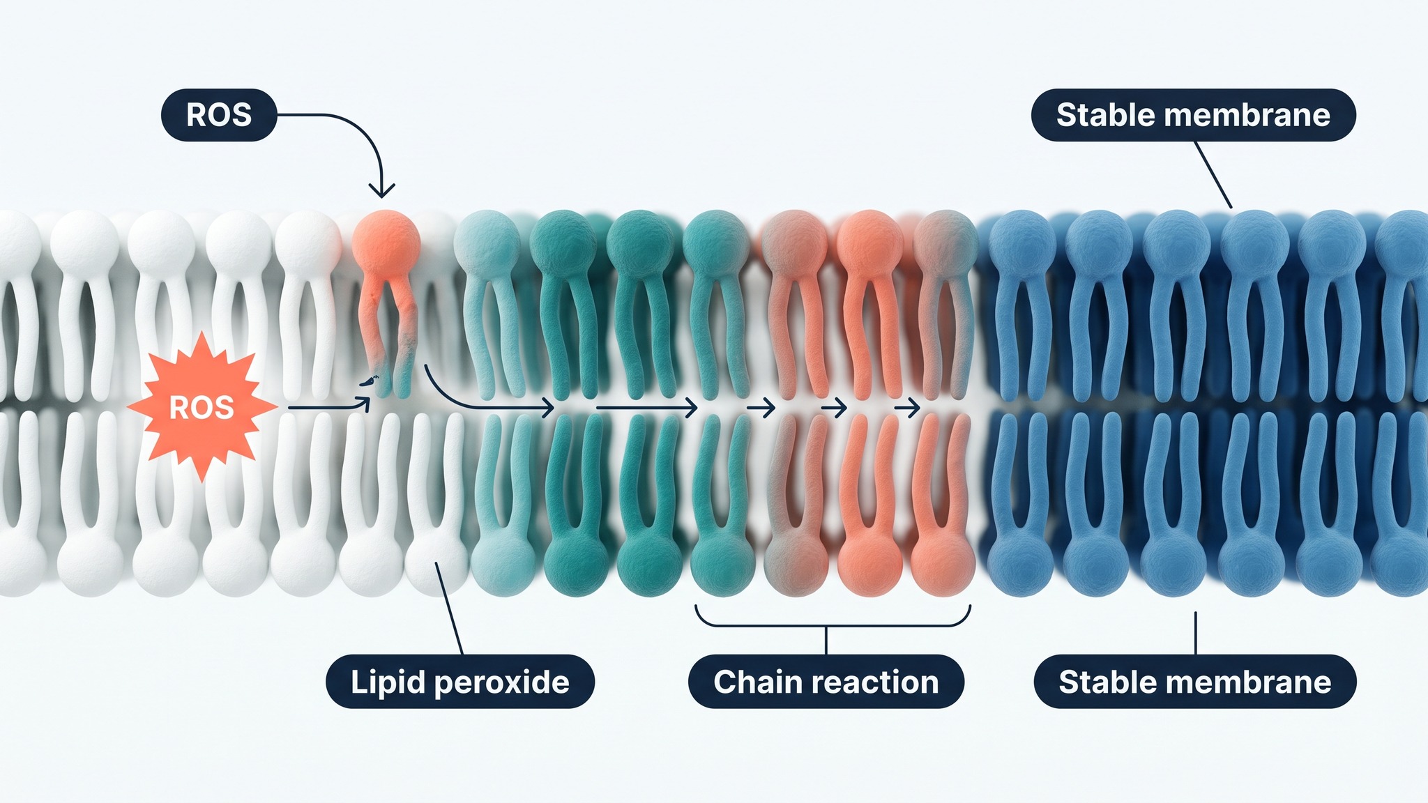
Glutathione does more than sit in a blood test. Inside a neuron, GSH helps control the redox environment around membranes, proteins, and mitochondrial enzymes. When the reserve falls, lipid peroxidation can spread through polyunsaturated fats in a membrane. Protein oxidation can change enzyme function. DNA oxidation can increase repair demand. Nrf2 is the transcription factor that helps cells turn on antioxidant-production genes, including genes that support glutathione synthesis and recycling. A small blood study in young children reported lower Nrf2, GSH, and glutathione reductase with higher GSSG in autism spectrum disorder.
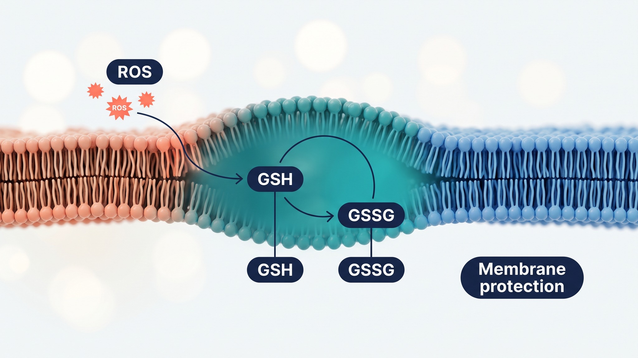
STUDY In autism cerebellum and BA22 tissue, lower GSH/GSSG occurred alongside higher protein and DNA oxidation markers, tying antioxidant capacity to damage measured directly in the brain. Rose et al., 2012, Evidence of oxidative damage and inflammation associated with low glutathione redox status in the autism brain, Translational Psychiatry.
How oxidative stress and ASD symptoms can track together
Oxidative stress in ASD can track with symptoms because redox balance affects cells that use a great deal of energy, including neurons in language, attention, and sensory networks. A single marker offers one part of the communication profile. A low antioxidant reserve may add biological pressure to systems that already need precise energy handling. Prefrontal and temporal networks help with language, social understanding, and the meaning of sounds. BA22 gives the laboratory story a concrete brain region to discuss.
Sensory overload has also been linked with measurable biology. In a study of 44 autistic children and 40 controls, higher blood 8-isoprostane and cysteinyl leukotrienes matched higher sensory sensitivity scores on the Short Sensory Profile. The finding is useful because 8-isoprostane reflects lipid peroxidation, while cysteinyl leukotrienes connect oxidative stress and neuroinflammation. These markers are research signals that require clinical context.
A newer 89-child study gives the same question a simpler frame. Children with autism spectrum disorder had higher 8-OHdG, 3-NT, and AOPP, plus lower OGG1, the enzyme that starts repair of oxidized DNA. Each of those findings tracked with higher clinician-rated DSM-5 symptom severity. The result describes a pattern of oxidative damage and lower repair capacity. Treatment choices still need the child’s complete clinical picture.
The 89-child oxidative biomarker study
Cohort composition. Ismael and Ismail, 2025, DOI 10.1007/s12031-025-02392-x. The study reported higher 8-OHdG, 3-NT, and AOPP, lower OGG1, and correlations with DSM-5 severity.
My child becomes much more sensitive to sound, sleep, and routine changes after an infection or a period of poor sleep. Could oxidative stress be part of that pattern, and which laboratory markers are worth discussing with a clinician?
Redox stress can be one piece of that pattern because infection, poor sleep, inflammation, and high energy demand can all shift redox balance. A clinician may look at the whole history first, then decide whether markers such as glutathione status, lipid-peroxidation markers, nutrient status, lactate, or carnitine fit the situation. The value comes from connecting results with symptoms, timing, medications, diet, and gastrointestinal health.
When symptoms and biology seem disconnected
Autism and Learning helps parents connect a child’s learning profile with the practical, biological questions that often sit behind difficult days. Buy the bookWhy repetitive behavior needs a careful evidence label
Repetitive and stereotypic behavior belongs in the article, with a clear boundary around the evidence. In a mouse model, lower antioxidant levels predicted more repetitive behavior. That is early-stage animal research. It helps researchers test mechanisms. Human studies are the next step before this finding can guide care for children.
STUDY In 44 autistic children, 8-isoprostane and cysteinyl leukotrienes were higher than in controls and both correlated strongly with Short Sensory Profile scores. Qasem et al., 2016, Cysteinyl leukotriene correlated with 8-isoprostane levels as predictive biomarkers for sensory dysfunction in autism, Lipids in Health and Disease.
How antioxidants in autism cover different parts of oxidative stress
Antioxidant support is often recommended because it can neutralize reactive damage or rebuild the antioxidant supply. N-acetylcysteine, or NAC, supplies cysteine for glutathione synthesis. Cysteine-rich whey protein aims at the same upstream need. CoQ10 supports electron transport, vitamin E helps protect lipid membranes, and polyphenols can influence redox-signaling pathways including Nrf2. Each target is different, so a supplement list is less useful than asking which part of the redox system appears under pressure.
The human evidence is mixed and specific. A randomized, double-blind study enrolled 46 preschool children with autism spectrum disorder in a 90-day cysteine-rich whey protein intervention. Forty completed it. The active group increased glutathione and showed a significant advantage in the VABS-II composite score. That result supports further study of glutathione precursors. It also gives families a reason to discuss measurement, dose, tolerance, food allergies, and the child's complete clinical picture with a qualified clinician.
| Marker or pathway | What it can describe in an oxidative-stress assessment |
|---|---|
| GSH, GSSG, GSH/GSSG | Reduced antioxidant reserve, oxidized glutathione burden, and redox balance. |
| 8-isoprostane and lipid peroxides | Oxidative injury to membrane fats, with 8-isoprostane studied in sensory dysfunction. |
| 8-OHdG, OGG1, 3-NT, AOPP | DNA oxidation, DNA-repair capacity, protein nitration, and oxidized protein products. |
| Lactate, carnitine, PCr/Cr, creatine-kinase activity | Exploratory energy-handling context that needs interpretation alongside symptoms and standard clinical assessment. |
Why creatine deserves its own place beside antioxidants
Creatine has a modest direct scavenger story and a more important energy story. Experimental work suggests creatine can react with some reactive oxygen and nitrogen species. That effect can help at the margin. The more distinctive mechanism is indirect. Creatine gives mitochondrial creatine kinase the substrate it needs to keep ADP moving back toward ATP production. A working ADP-recycling loop reduces the electron backlog that favors mitochondrial ROS. In this model, creatine is less like a sponge for damage and more like a way to prevent some damage from being generated. For a fuller clinical overview, see Is creatine good for autistic people?
STUDY In a randomized trial of cysteine-rich whey protein, children receiving the intervention increased glutathione and showed significant VABS-II composite-score improvement versus placebo. Castejon et al., 2021, Improving Antioxidant Capacity in Children With Autism, Frontiers in Psychiatry.
How mitochondrial creatine kinase may reduce oxidative stress at its source
Mitochondrial creatine kinase is an enzyme in the mitochondrial intermembrane space. It converts creatine and ATP into phosphocreatine and ADP. The newly formed ADP can return to the mitochondrial matrix and keep ATP synthase working. This is ADP recycling. It matters because a respiratory chain with an adequate ADP sink is less likely to remain in the highly charged state that favors electron leak and ROS formation. The system therefore addresses oxidative stress upstream, before a membrane lipid or protein needs to be repaired.
Antioxidant cleanup
NAC, glutathione precursors, vitamin E, CoQ10, and polyphenols can support antioxidant capacity or neutralize reactive damage after ROS has appeared.
Creatine kinase and ADP recycling
Creatine supports the phosphocreatine system. Mitochondrial creatine kinase can recycle ADP, sustain respiration, and lower ROS generation in isolated brain mitochondria and cortical-neuron experiments.
The phosphocreatine-to-creatine ratio, written PCr/Cr, changes how strongly this effect appears in the laboratory. It provides an energetic-state readout for research. In the 2006 mitochondrial creatine-kinase study, creatine activation produced state-3-like respiration in isolated brain mitochondria and reduced hydrogen-peroxide generation through enzyme-dependent ADP recycling. The work also tested embryonic rat cortical neurons. The core mechanism is decades-established neuroscience and mitochondrial biology. Autism-specific treatment studies are the next needed step.
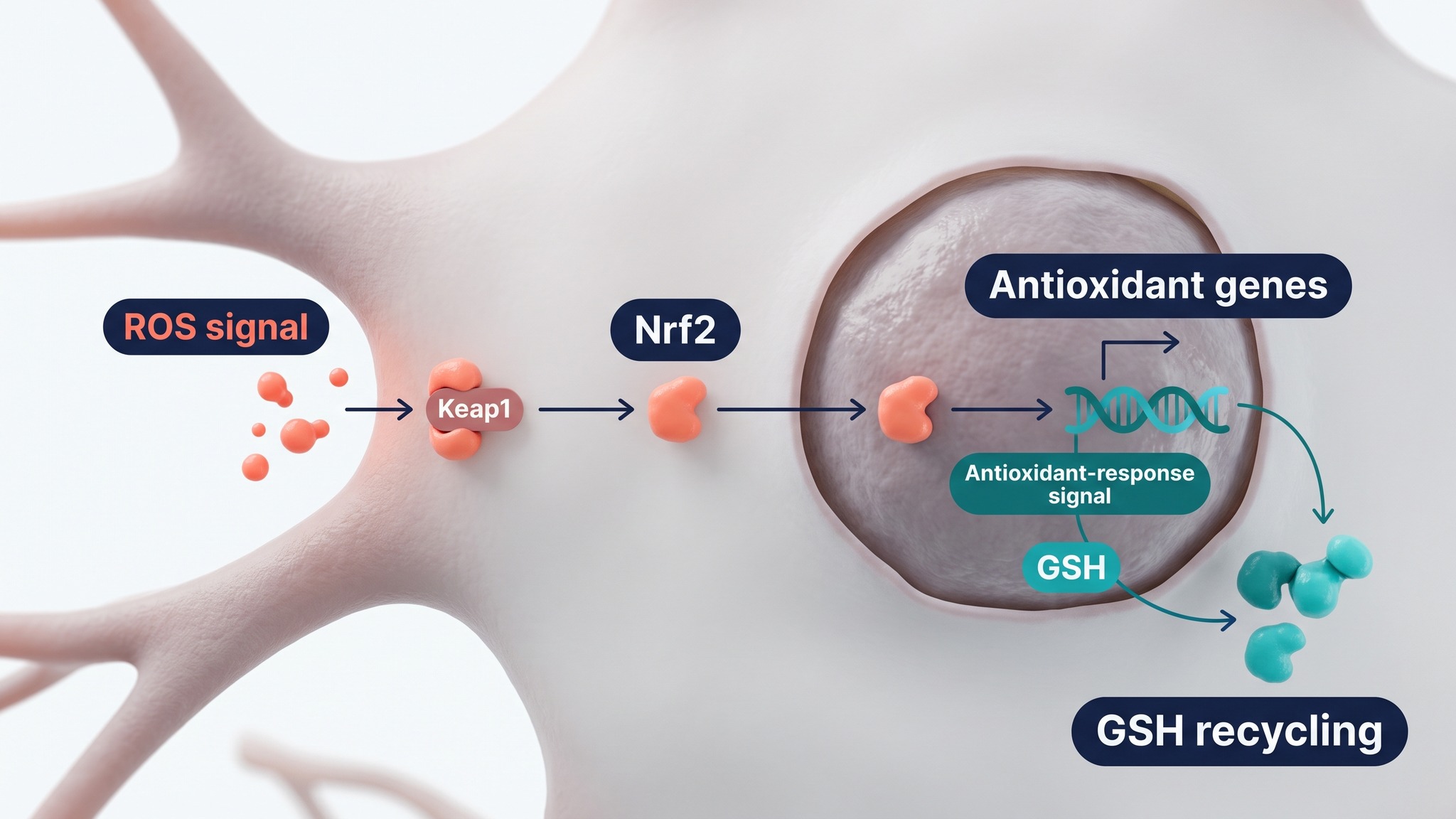
Mitochondrial kinase activity performed a key role as a preventive antioxidant against oxidative stress.
STUDY In isolated rat brain mitochondria and embryonic cortical neurons, mitochondrial creatine-kinase activity reduced hydrogen-peroxide and ROS generation through ADP recycling, with the effect dependent on the PCr/Cr ratio. Meyer et al., 2006, Mitochondrial creatine kinase activity prevents reactive oxygen species generation, Journal of Biological Chemistry.
Why mitochondrial dysfunction and oxidative stress in autism belong in one story
Mitochondrial strain and redox imbalance are closely connected because mitochondria make ATP and can also become a major source of ROS. This is the link between oxidative stress and mitochondrial dysfunction. A ³¹P-MRS scan measures energy molecules without a biopsy. Small autism studies have reported lower brain phosphocreatine or trends in that direction, especially in children selected for suspected mitochondrial dysfunction. Creatine sits at the intersection. It buffers ATP demand through phosphocreatine, and its kinase system can keep ADP available to the mitochondrial machinery. The related creatine deficiency and autism guide explains the diagnostic pathway in more detail.
Propionic acid adds a gut-mitochondria route to the story. It is a normal short-chain fatty acid made by the microbiome and used in human metabolism. In autism-derived lymphoblastoid cell lines, its mitochondrial effect depended on concentration, exposure time, and redox state. Added ROS removed the benefit seen at some propionic-acid exposures and reduced reserve capacity. A stool result represents one layer of a wider gut-mitochondria hypothesis. It shows why microbiome metabolites, redox status, and mitochondrial function are reasonable to study together.
What a small ³¹P-MRS autism study observed
Golomb et al., 2014, DOI 10.1177/0883073813498466. Six ASD cases with suspected mitochondrial dysfunction and six matched controls. These are preliminary findings.
Children can have very different mitochondrial and redox profiles. Energy strain can help explain why sleep loss, infection, gastrointestinal symptoms, restricted intake, and rapid developmental demands can make some days much harder. A clinician who sees a pattern may consider whether oxidative stress biomarkers, antioxidant enzyme activities, carnitine metabolism, lactate, and the creatine-phosphocreatine system deserve a more complete evaluation.
STUDY A preliminary ³¹P-MRS case-control study found lower intracellular muscle pH in all six ASD cases, with brain phosphocreatine trends in three of four scanned cases. Golomb et al., 2014, Assessing bioenergetic compromise in autism spectrum disorder with 31P magnetic resonance spectroscopy, Journal of Child Neurology.
STUDY In autism-derived lymphoblastoid cell lines, propionic acid altered mitochondrial function according to concentration, duration, and redox state, while experimentally increased ROS reduced reserve capacity. Frye et al., 2016, Modulation of mitochondrial function by the microbiome metabolite propionic acid in autism and control cell lines, Translational Psychiatry.
What oxidative stress means for your child now
Oxidative stress becomes useful in practice when it turns a confusing group of symptoms and laboratory results into a focused clinical conversation. Low glutathione, weak mitochondrial energy, and altered creatine handling can each offer a practical starting point for investigating an individual autistic child’s pattern.
Start with the pattern you see at home
Bring the child’s timeline to the appointment. Include sleep, illness, regression or plateaus, sensory overload, diet, bowel symptoms, medications, supplements, growth, fatigue, and every laboratory result. That turns a vague conversation about stress in autism spectrum disorder into a focused search for sources of energy strain and oxidative load. It also protects families from chasing a single supplement because a laboratory value was flagged. If a previously established skill has been lost, the skills regression and autism guide offers a focused way to map that change.
A systematic review and meta-analysis of oxidative stress marker aberrations in children with autism spectrum disorder describes a broad research field with varied profiles. Autism spectrum disorder is a neurodevelopmental disorder with substantial biological variation. The role of oxidative stress may differ with age, diet, infection burden, sleep, medication, and mitochondrial dysfunction in autism spectrum. The pattern can include dysfunction in autism spectrum disorder. That is why oxidative stress markers, markers of oxidative stress, oxidative stress and antioxidant measures, antioxidant enzyme activities, and an oxidative stress status are best interpreted as part of metabolism in autism spectrum disorder. This approach also makes room for metabolism and oxidative stress, neuroinflammation in autism, and neuroinflammation alongside oxidative stress within research on the diagnosis of autism spectrum disorder.
Why creatine belongs in the conversation
The creatine-kinase mechanism is already well described in neuroscience and mitochondrial research, including Parkinson’s and other neurodegenerative work. Autism is the frontier. The question is whether children with a demonstrable energy and redox pattern could benefit from a pathway-specific approach that joins creatine handling, mitochondrial function, glutathione support, and the factors that keep increasing ROS. That is a sharper question than asking whether one antioxidant works for every child.
Research language can sound more certain than a family’s lived experience. Increased oxidative stress, oxidative stress in children, and oxidative stress in the brain are biological signals related to oxidative stress that need clinical context. The levels of oxidative stress can change with context. Some studies use the Childhood Autism Rating Scale when discussing autism severity. Others examine activity in children with autism, irritability in children with autism, or stress in children with autism. A rat model of autism can test whether a pathway may cause oxidative stress, while children remain vulnerable to oxidative stress or susceptible to oxidative stress for many different reasons. Researchers also discuss oxidative stress in individuals, stress in individuals with ASD, neuroinflammation and oxidative stress, reducing oxidative stress, and linking oxidative pathways. These terms support screening of autism spectrum disorder and biomarkers for autism spectrum disorder research, while clinical care stays anchored to the child in front of you.
My child has low glutathione and borderline lactate or carnitine results, but the biggest concern is speech and sensory overload. Could creatine, phosphocreatine, and mitochondrial energy help explain why these symptoms fluctuate?
They may help frame a useful clinical question. Creatine and phosphocreatine buffer short bursts of energy demand, while mitochondrial creatine kinase helps recycle ADP for ATP production. If a child also has low glutathione or signs of oxidative stress, the energy system may face extra pressure during illness, poor sleep, or rapid growth. This research field calls for a clinician-led review of the full pattern before any supplement plan is chosen.
Make the biology of learning easier to follow
Autism and Learning gives parents a practical framework for linking learning, energy demands, symptoms, and the questions worth taking to the next appointment.
Frequently asked questions
Common research markers include reduced and oxidized glutathione, the GSH/GSSG ratio, 8-isoprostane, malondialdehyde, 8-OHdG, 3-nitrotyrosine, AOPP, and antioxidant enzyme activities. Nrf2 and glutathione-recycling enzymes describe the antioxidant response. Lactate, carnitine, phosphocreatine-to-creatine ratio, and creatine-kinase activity add exploratory energy-metabolism context that needs clinical interpretation.
Speech and language depend on high-energy cortical networks, including temporal regions that process speech and prefrontal networks that organize attention and social meaning. A plausible biochemical route begins with mitochondrial ROS. ROS can consume GSH, increase lipid peroxidation, and alter membrane and mitochondrial function. Postmortem BA22 findings show lower glutathione redox capacity with higher protein and DNA oxidation. These data support a biological link worth studying, while each child’s language profile still reflects many developmental and environmental factors.
Mitochondria produce ATP and can also generate ROS. When energy handling is strained, mitochondrial dysfunction and oxidative stress can reinforce each other. 31P-MRS studies have reported preliminary phosphocreatine differences in selected autistic children. Propionic acid, a microbiome-derived short-chain fatty acid, changed mitochondrial performance in autism-derived cell lines according to dose, exposure time, and redox state. Creatine and phosphocreatine sit at this intersection because they buffer energy demand and support ADP recycling through creatine kinase.