IBS and mast cells: why the gut barrier stays on edge
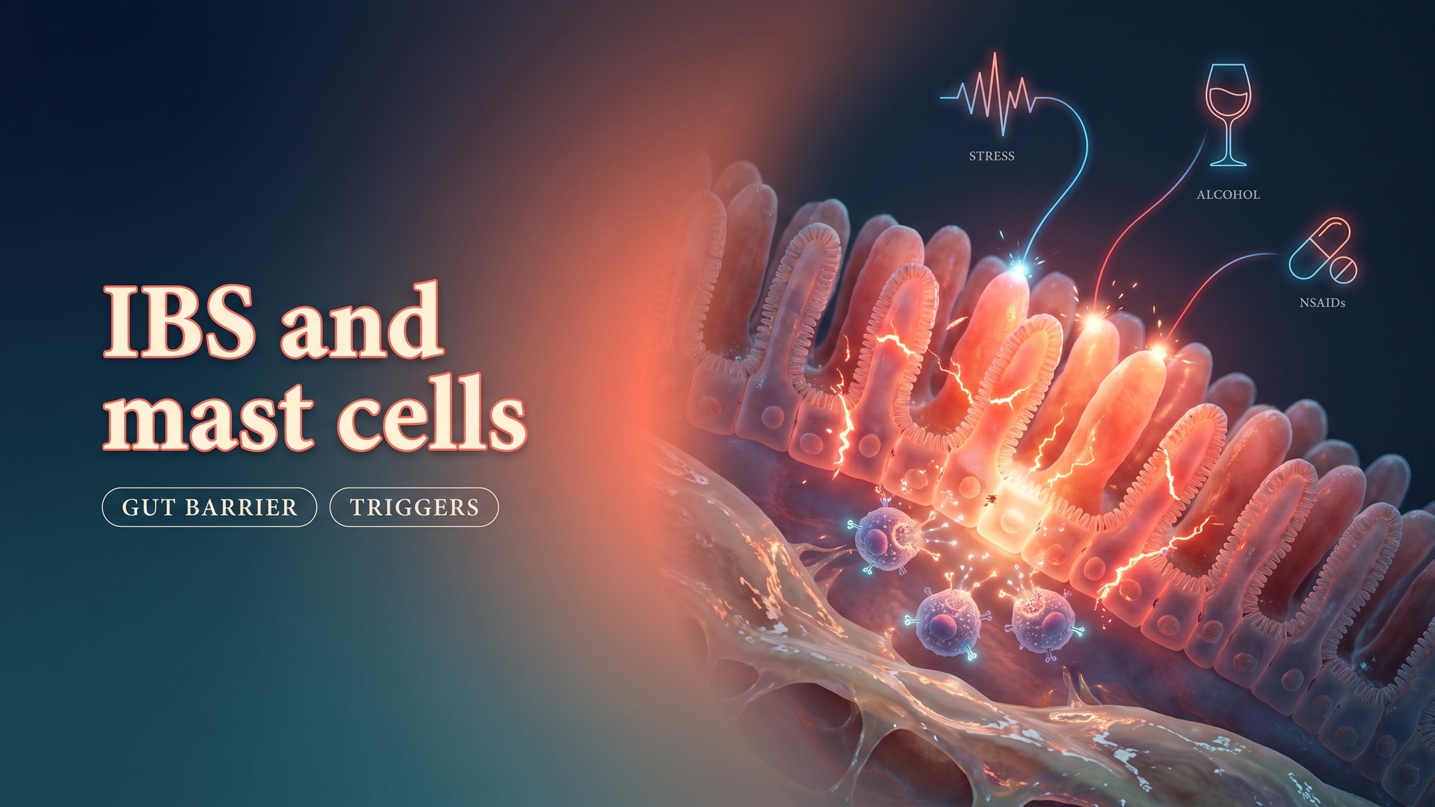
IBS and mast cells meet at the intestinal barrier. A leakier mucosa, reactive immune cells, microbial signals and familiar daily triggers can keep the gut in a state of heightened response. The same biology also reveals practical routes toward a calmer barrier through butyrate, quercetin and linden tea.
What is the link between IBS and mast cells?
The connection between IBS and mast cells becomes clearer when prevalence and location are considered together. Irritable bowel syndrome affects roughly 10 percent of the population, while mast cell disorders are estimated at 1 to 4 percent. Within that overlap, mast cell activation syndrome stands out. Its prevalence among people with IBS is reported as 16.3 times higher than in the general population. This places mast cell activity within the clinical reality of IBS rather than at the edge of it.
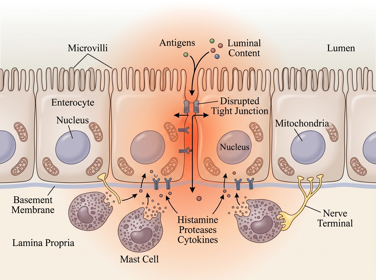
Mast cells are positioned close to the intestinal barrier, where they can respond rapidly to stress signals, microbial products and chemical exposures. When that response becomes excessive, the gut can become more permeable and more sensitive. This creates a plausible biological route from immune activation to urgency, loose stools, abdominal discomfort and a bowel that reacts strongly to otherwise ordinary inputs.
Relative prevalence of mast cell activation syndrome in IBS
Data source: DOI 10.1111/nmo.14265
STUDY Mast cell activation syndrome was reported 16.3 times more frequently among people with IBS than in the general population. Source study, DOI 10.1111/nmo.14265.
Where are mast cells increased in the gut?
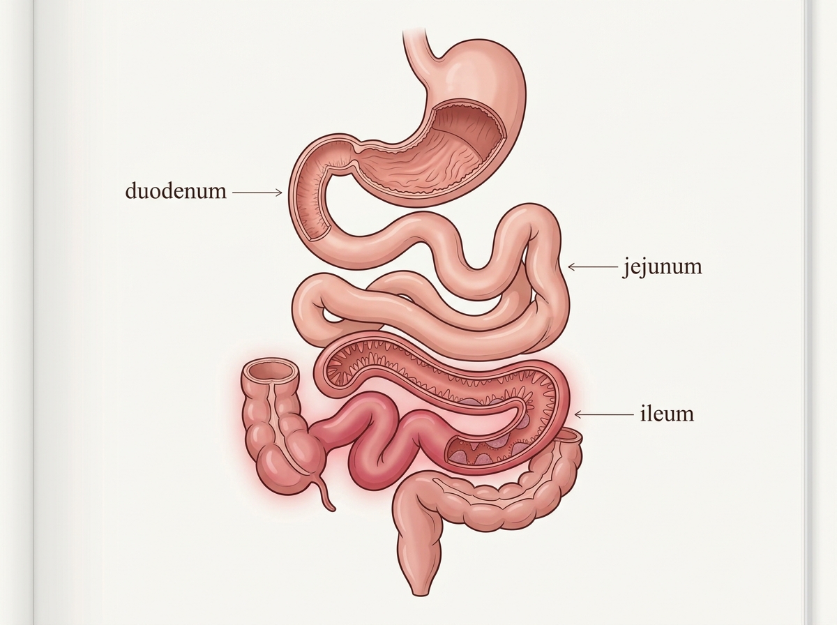
Mast cells are present across the small intestine, yet the ileum shows the clearest increase in IBS. The standardized mean difference is 1.78 in the ileum, compared with 0.81 in the duodenum and 0.58 in the jejunum. The pattern is therefore uneven. The strongest enrichment appears in the distal part of the small intestine.
This anatomical distribution matters because it points to a specific region where immune-cell density may contribute more strongly to symptom generation. A higher number of mast cells close to the mucosa increases the opportunity for local mediators to influence the barrier. The ileum therefore becomes a key part of the IBS picture when mast cell reactivity and intestinal permeability are both prominent.
Mast cell density by small-bowel segment in IBS
Data source: DOI 10.1111/nmo.13718
STUDY The ileum showed the largest increase in mast cell density, ahead of both the duodenum and jejunum. Source study, DOI 10.1111/nmo.13718.
How do mast cells affect intestinal permeability?
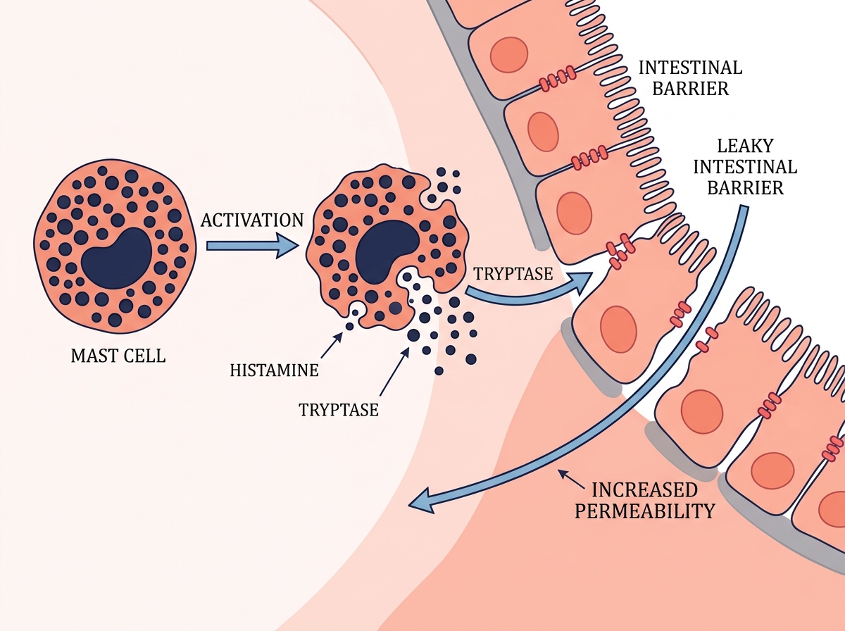
Mast cell activity and intestinal permeability rise together in diarrhea-predominant IBS. Tryptase is reported at 0.86 in IBS-D compared with 0.28 in healthy controls. Permeability reaches 0.644 compared with 0.06. The scale of the difference places the intestinal barrier at the center of the mechanism.
Tryptase is released during mast cell activation. As mast cell activity increases, the barrier becomes easier to cross. More material from the intestinal lumen can then reach the immune-facing side of the mucosa, creating additional stimulation. This feedback pattern helps explain why the bowel can remain reactive after the original trigger has passed and why IBS-D often feels like a system that has lost its normal threshold.
Tryptase and permeability in IBS-D versus healthy controls
Data source: DOI 10.5056/jnm.2013.19.2.244. Each metric pair is scaled independently.
STUDY Higher mucosal tryptase was reported alongside markedly greater intestinal permeability in IBS-D. Source study, DOI 10.5056/jnm.2013.19.2.244.
Need a more personalized gut map?
If your IBS picture includes food reactivity, barrier symptoms or a mast cell pattern, we can investigate the physiology together and build a more targeted plan.
Which habits and exposures push mast cells to flare?
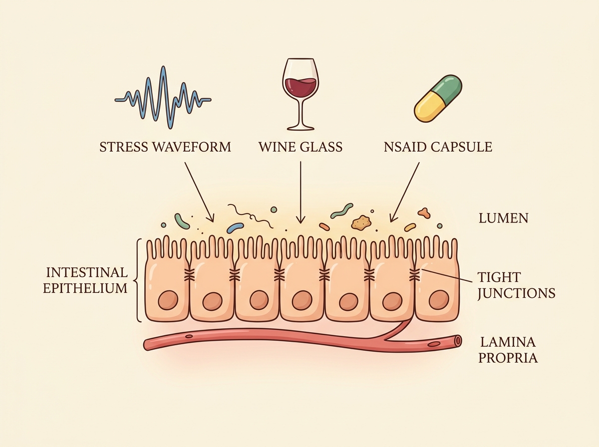
Chronic stress, alcohol and NSAIDs reach the intestinal barrier through different mechanisms, yet all three can intensify mast cell reactivity. Chronic stress activates CRF-linked pathways and increases permeability, a pattern associated with visceral hypersensitivity. Alcohol is converted to acetaldehyde, which promotes mast cell degranulation and is linked to a 45 to 59 percent rise in permeability. NSAIDs such as diclofenac activate PAR-2 and reduce tight-junction quality.
The practical consequence is cumulative. A stressful period, repeated alcohol exposure and frequent NSAID use can all load pressure onto the same barrier system. The result is a gut that opens more easily, reacts more intensely and takes longer to return to baseline. Recognizing these mechanisms gives recurring symptom flares a clearer structure.
| Trigger | Mechanism |
|---|---|
| Chronic stress | CRF activation → increased permeability |
| Alcohol | Acetaldehyde-driven mast cell activation → 45–59% rise in permeability |
| NSAIDs | PAR-2 activation → tight-junction impairment |
STUDY Chronic stress, alcohol and NSAIDs reach the intestinal barrier through distinct pathways that converge on mast cell activation and barrier disruption. Stress source, DOI 10.1007/s10753-021-01424-z. Alcohol source, DOI 10.1016/j.bbi.2014.12.010. NSAID source, DOI 10.1002/jcph.737.
Which microbiome patterns make mast cell-related IBS worse?
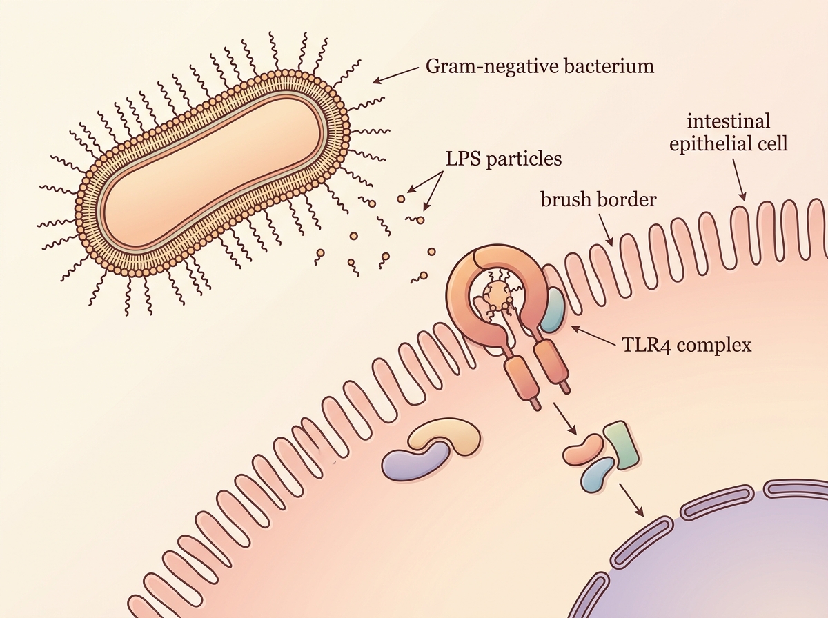
A dysbiotic microbiome can intensify mast cell-related IBS when Gram-negative, LPS-producing bacteria become more prominent. LPS binds to TLR4 and activates inflammatory signaling at the intestinal surface. As this pressure continues, barrier integrity declines, diarrheal symptoms become more likely and the intestinal environment becomes easier for pathogens to exploit.
The important feature is the direction of the microbial output. A microbiome dominated by LPS-rich signals continually sends activation messages toward the barrier. Mast cells receive more stimulation, epithelial control weakens and the gut becomes less resilient. This links dysbiosis directly with immune activation rather than treating the microbiome as a separate issue.
| Bacterial pattern | Mechanism |
|---|---|
| Gram-negative LPS+ bacteria | TLR4 binding |
| Pathogen expansion | Higher luminal LPS load |
| Functional outcome | Barrier loss and diarrheal tendency |
STUDY Gram-negative bacterial expansion can increase LPS-TLR4 signaling and contribute to intestinal barrier injury. Source study, DOI 10.1172/jci.insight.146529.
Which microbiome patterns help protect the gut barrier?
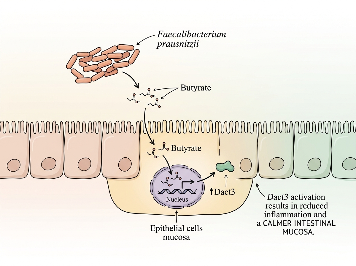
The protective microbiome pathway begins with Faecalibacterium prausnitzii. This bacterium produces butyrate, and butyrate promotes Dact3 expression in the intestinal mucosa. The sequence supports anti-inflammatory signaling and helps protect the intestinal wall from excessive mast cell activity.
This pathway shifts the focus from bacterial names alone to what those bacteria produce. Butyrate is the functional bridge. It connects a protective microbial pattern with a mucosa that is better equipped to regulate inflammation. When that route is supported, the intestinal barrier faces fewer signals that keep mast cells in a persistently activated state.
STUDY Faecalibacterium prausnitzii, butyrate and Dact3 form a protective pathway that supports intestinal homeostasis and moderates mast cell-driven inflammation. Source study, DOI 10.1080/19490976.2020.1826748.
Can quercetin help stabilize mast cells?
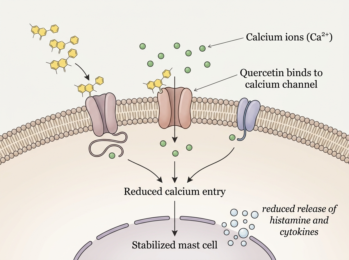
Quercetin is a naturally occurring flavonoid with mast cell-stabilizing activity. Its mechanism includes inhibition of calcium channels in mast cells and modulation of cytokine activity. Because calcium entry is central to degranulation, reducing that signal can make mast cells less likely to release their inflammatory contents.
Quercetin can be obtained through foods and herbs, which makes it relevant to a practical gut-barrier strategy. Its value lies in the combination of accessibility and mechanism. It acts at the level of mast cell activation while also fitting into a broader dietary pattern built around flavonoid-rich plant compounds.
STUDY Quercetin can stabilize mast cells by moderating calcium-dependent activation and inflammatory cytokine signaling. Source study, DOI 10.3389/fimmu.2024.1418897.
How do you prepare linden tea in a way that fits this gut-barrier framework?
Linden flowers provide a practical herbal source of quercetin and other flavonoids. Their phytochemical profile includes 42 identified flavonoids, with 21 quantified compounds. This breadth makes linden tea a natural fit for a routine centered on mast cell stability and a calmer intestinal barrier.
The ideal preparation uses 1.5 g of dried linden flowers in 150 ml of near-boiling water. The infusion steeps for 5 to 15 minutes and is then strained. The referenced use is 2 to 4 times daily. A measured dose, controlled water volume and sufficient steeping time create a more consistent preparation than a loosely filled cup.
Flavonoid profile highlighted for linden flowers
Data source: DOI 10.1016/j.indcrop.2020.112691
STUDY Linden flowers contain a broad range of flavonoids, including quercetin-related compounds. Source study, DOI 10.1016/j.indcrop.2020.112691.
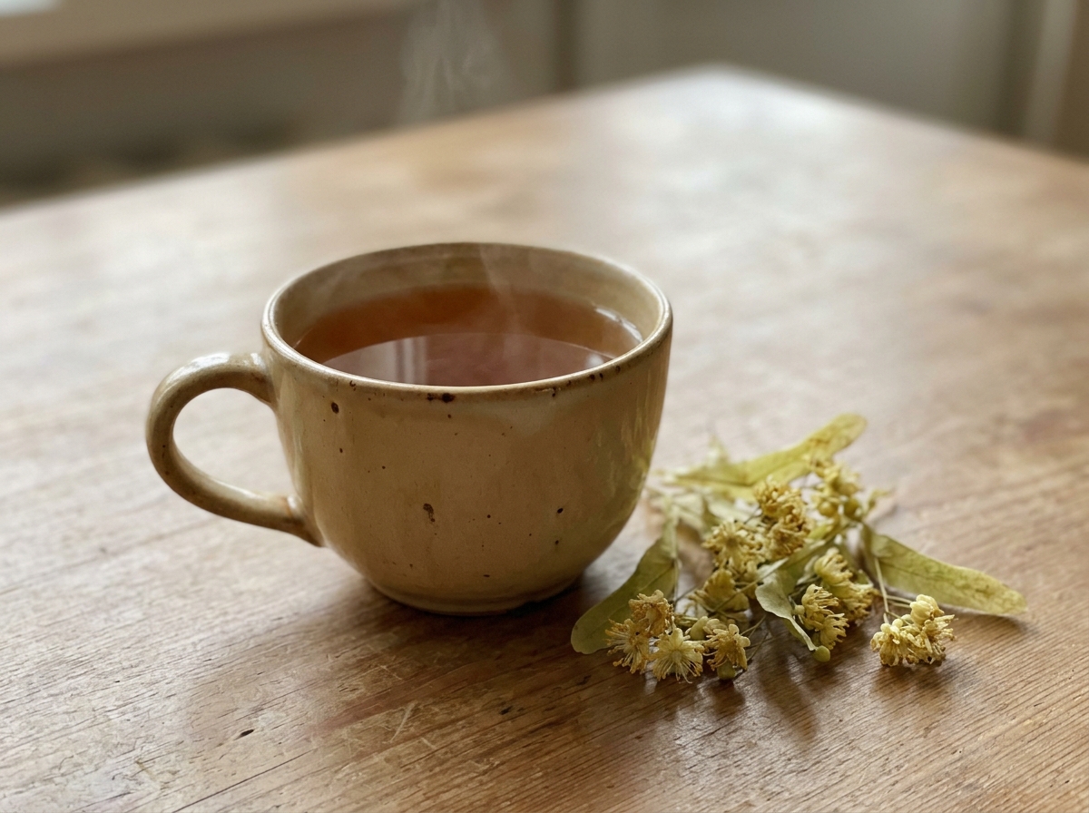
Linden tea
Ingredients
- 1.5 g dried linden flowers
- 150 ml near-boiling water
Method
- Place the dried linden flowers in a cup or teapot.
- Pour in 150 ml of near-boiling water.
- Let the infusion steep for 5 to 15 minutes.
- Strain and drink warm.
- Use 2 to 4 times daily when this preparation fits the wider clinical context.
Need a more personalized gut map?
If your IBS picture includes food reactivity, barrier symptoms or a mast cell pattern, we can investigate the physiology together and build a more targeted plan.
IBSyncrasy
IBSyncrasy is my practical book on irritable bowel syndrome. It connects symptom patterns with physiology, testing logic and everyday interventions, so complex gut stories become easier to read and act on.
Buy IBSyncrasyFrequently asked questions
Mast cells are especially relevant in the IBS subgroup marked by intestinal permeability, diarrheal symptoms, trigger sensitivity and immune reactivity.
The ileum shows the strongest increase in mast cell density among the small-bowel segments compared, making it a key anatomical site in this mechanism.
Gram-negative, LPS-producing patterns activate TLR4, weaken the barrier and maintain inflammatory signaling close to mast cells.
Linden flowers contain quercetin and a broad flavonoid profile, making the infusion a practical botanical source within this framework.